X-Ray Elastography Images Soft-Tissue Tumors
By MedImaging International staff writers
Posted on 16 Apr 2020
A new study suggests that elastography can be adapted to X-ray imaging of soft tissues to reveal tumors earlier than other techniques.Posted on 16 Apr 2020
Developed by researchers at Tohoku University (Japan), the Institute of Materials Structure Science (IMSS-KEK; Ibaraki, Japan), and other institutions, dynamic X-ray elastography is a novel imaging modality that relies on shear-wave propagation to map elastic modulus in soft viscoelastic materials, which has far only been possible via magnetic resonance imaging (MRI) and ultrasound imaging. However, X-rays can provide a thousand-fold greater resolution than ultrasound.

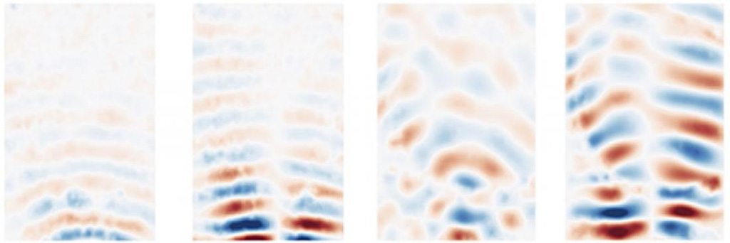
Image: X-ray elastography images showing the stiffness of different materials (Photo courtesy of Wataru Yashiro/ Tohoku University)
For the study, the researchers pneumatically vibrated polyacrylamide gel phantoms to obtain two-dimensional (2D) maps of storage and loss moduli, which were determined from the temporal variation in displacement vectors of the vibrating particles in the phantom models. The results revealed a storage moduli precision of 30% and a spatial resolution several orders of magnitude higher than that of MRI or ultrasound imaging modalities. The study was published in the April 2020 issue of Applied Physics Express.
“The resulting images would be much higher resolution, able to spot things on the scale of microns instead of millimeters,” said senior author Wataru Yashiro, PhD, of Tohoku University. “This greater precision doesn't just mean identification of much smaller or deeper lesions, but, importantly for patients, because smaller lesions can be newer ones, potentially also much earlier on in a disease or condition.”
Elastography is a medical imaging modality that maps the elastic properties and stiffness of soft tissues in order to provide diagnostic information. The technique works by creating a distortion in the tissue, observing the tissue response to infer the mechanical properties of the tissue, and then display the results as an image. The most prominent techniques use ultrasound or MRI to make both the stiffness map and an anatomical image for comparison.
Related Links:
Tohoku University
Institute of Materials Structure Science


