Obese Patients Are Exposed to Higher Radiation Doses
By MedImaging International staff writers
Posted on 10 Jan 2019
A new study finds that obese patients face an increased radiation-related lifetime cancer risk due to higher dose area product (DAP) radiation exposures.Posted on 10 Jan 2019
Researchers at the University of Exeter (United Kingdom), Musgrove Park Hospital (Taunton, United Kingdom), and Derriford Hospital (Plymouth, United Kingdom) conducted a study involving 630 patients from a bariatric clinic who had a projection radiography history. The prime purpose of the study was to evaluate X-ray radiation dose delivered to obese patients, and its relationship to patient size. A secondary purpose was to estimate subsequent projected radiation-related lifetime cancer risk in patients with obesity.

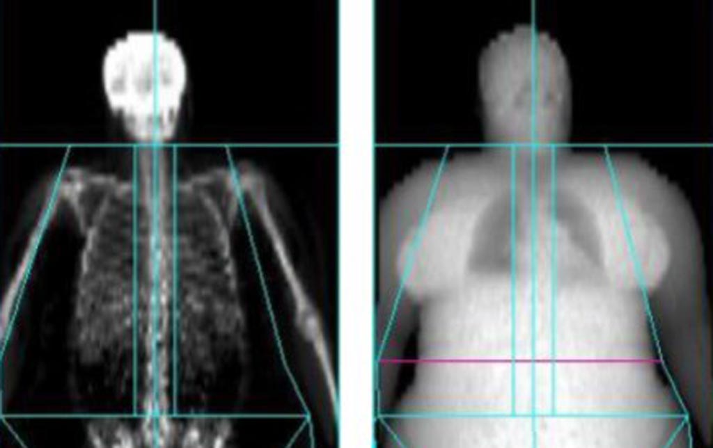
Image: A new study claims that obese patients are exposed to more radiation (Photo courtesy of University of Exeter).
Patients' DAP data were collected for all projection radiography, and the effective risk for patients with obesity was then compared to that of normal-weight patients. The results showed that patients with obesity received higher DAPs for all examinations, with the highest DAPs in abdominal and lumbar spine radiographs. In abdomen and chest x-rays, there were moderate-to-low correlations between patient size and DAP. The projected radiation-related lifetime cancer risk was up to 153% higher than in normal-weight adult patients. The study was published on December 20, 2018, in the Journal of Radiological Protection.
“As a researcher and a radiographer, I believe these radiation doses figures are only to be expected due to the lack of guidelines to aid imaging this group of patients,” said lead author Saeed Alqahtani, MD, of the University of Exeter, and colleagues. “As well as the doses of radiation given to the patient, many technical factors contribute to the image quality of an X-ray. We already started working on this issue in an attempt to produce prediction models that can aid radiographers to choose the best technical factors based on the patient's size.”
DAP reflects not only the dose within the radiation field but also the area of tissue irradiated. Therefore, it may be a better indicator of the overall risk of inducing cancer than the dose within the field. It also has the advantages of being easily measured, with the permanent installation of a DAP meter on the X-ray set. Due to the divergence of a beam emitted from a point source, the area irradiated increases with the square of distance from the source, while radiation intensity decreases according to the inverse square of distance. Consequently, the product of intensity and area, and therefore DAP, is independent of distance from the source.
Related Links:
University of Exeter
Musgrove Park Hospital
Derriford Hospital


