CT Scans Help Improve Hip Fracture Outcomes
By MedImaging International staff writers
Posted on 13 Jun 2017
The results of a new study suggest that clinicians could use CT scans to help find the best treatment for elderly patients who suffer from broken hips.Posted on 13 Jun 2017
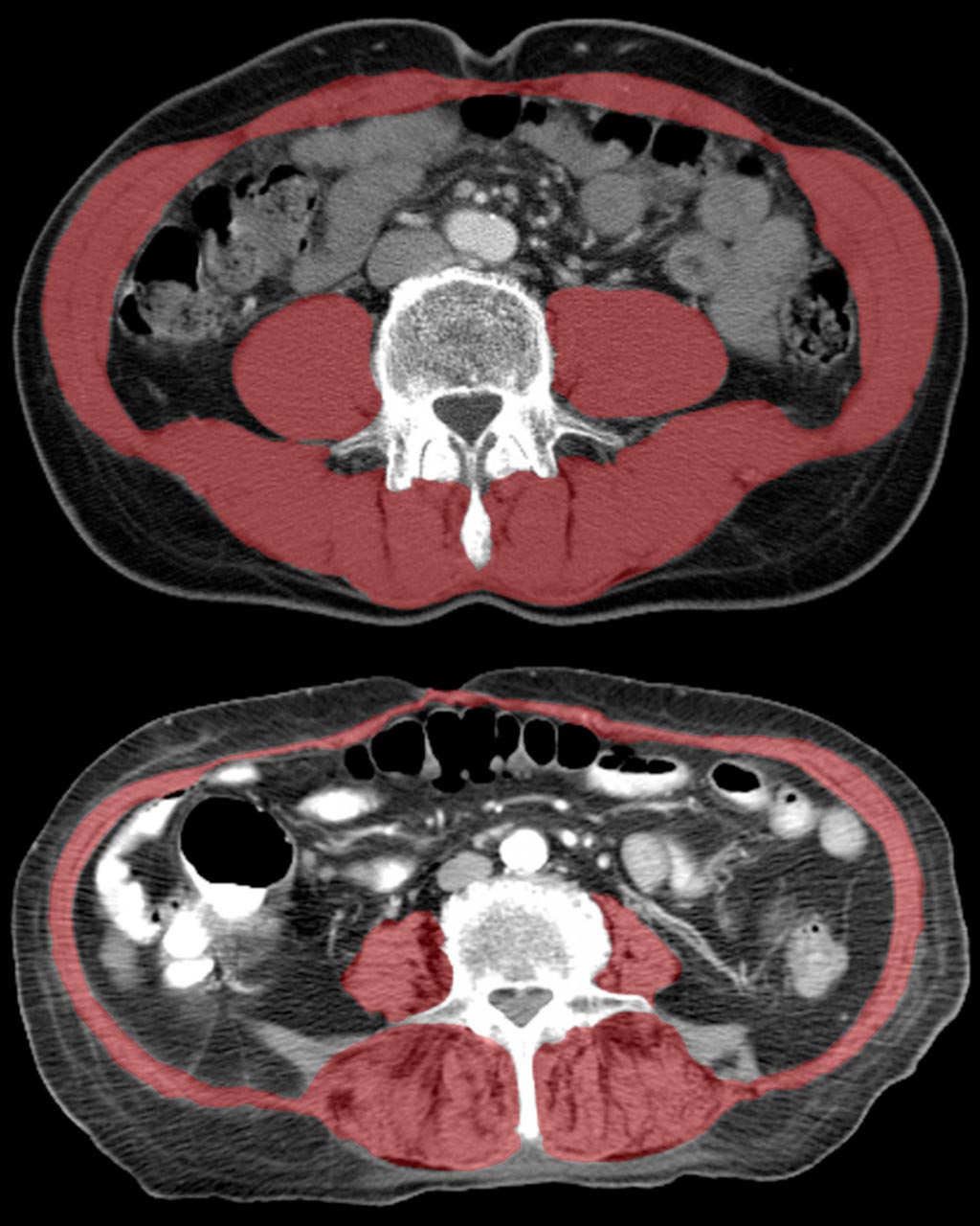
The researchers found that a decrease in the strength of the "core" muscle that stabilizes the spine of patients with hip fractures was related to a decrease in survival times of the patients.

Image: A new study of CTs shows that reduced size and density of core muscle (highlighted in red) as shown in the lower image is associated with frailty and reduced lifespan in hip-fracture patients (Photo courtesy of the University of California – Davis Health System).
The study was led by radiologists from UC Davis Medical Center (Sacramento, CA, USA) and the Wake Forest Baptist Medical Center (Winston-Salem, NC, USA), and the results were published in the June 2017 issue of the American Journal of Roentgenology. This study is the first to use Computed Tomography (CT) to specifically link hip fractures with survival of patients.
The study included nearly 300 patients, aged 65 years and over, who had been treated at UC Davis between 2005 and 2015, for suspected hip fractures. All patients underwent a CT exam to diagnose or rule out hip fractures. The researchers evaluated the size and density of the spinal lumbar and thoracic muscles and compared this data with mortality data. The researchers found that survival rates during the 10-year study were significantly higher for patients with better core muscle strength.
Lead author of the study, UC Davis professor of radiology, Robert Boutin, said, "As patients age, it becomes increasingly important to identify the safest and most beneficial orthopedic treatments, but there currently is no objective way to do this. Using CT scans to evaluate muscles in addition to hip bones can help predict longevity and personalize treatment to a patient's needs. We're excited because information on muscle is included on every routine CT scan of the chest, abdomen and pelvis, so the additional evaluations can be done without the costs of additional tests, equipment or software. Recognizing sarcopenia as a distinct condition that provides clues to future health can open doors to new discoveries in diagnosis and treatment."
Related Links:
UC Davis Medical Center
Wake Forest Baptist Medical Center


 Guided Devices.jpg)


