Mobile Phone Analysis Helps Reveal Radiation Exposure
By MedImaging International staff writers
Posted on 08 Jun 2017
An analysis of mobile phones and other objects in close proximity with the body could be used in retrospective dosimetry, according to a new study.Posted on 08 Jun 2017
Researchers at Lund University (Sweden) conducted a study that examined a number of objects and materials held close the body, and which therefore have a potential to provide information on whether the carrier has been exposed to ionizing radiation in the absence of dosimeters. Among the objects examined were mobile phones, teeth and dental fillings, household salt, and desiccant drying agents. The study showed that several of the materials contained very promising properties, not least of them mobile phones.

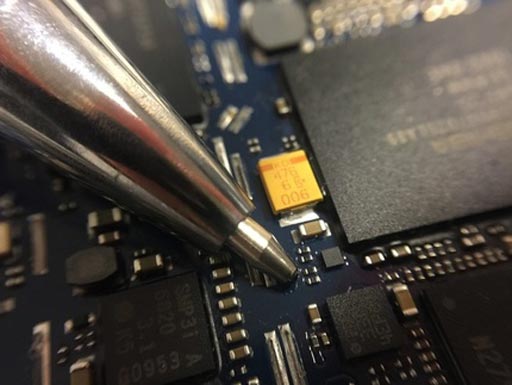
Image: A new study indicates mobile phone resistors could be used to retrospectively measure radiation dosage (Photo courtesy of Lund University).
Mobile phones contain resistors made from aluminum oxide (AlO), which can provide information about radiation as late as six years after the time of exposure. During analysis, the phone is dismantled and the resistor is examined using a light-sensitive measuring technique called optically stimulated luminescence (OSL), with results in two hours. The researchers also developed initial estimations of conversion factors for the transition between radiation dose and dosemeter material using an anthropomorphic phantom. The study was presented as a doctoral thesis at Lund University.
“In case of a nuclear power plant disaster, many people are worried, even when only a small number of people have been exposed to harmful levels of radiation,” said lead author medical physicist Therése Geber-Bergstrand, MSc, a doctoral student at Lund University. “The results from the mobile phones were very promising. Even though further studies are required, the phones can be used right away.”
OSL involves the stimulation of trapped electrons performed using light of a specific wavelength. The stimulation with light can be performed using different modes: continuous wave (CW-OSL), linearly modulated (LM-OSL), and pulsed (POSL). The CW-OSL mode is the most frequently used, with the intensity of the stimulation light kept constant and the luminescence recorded during stimulation. This involves using filters to discriminate between the stimulation light and the luminescence.
Related Links:
Lund University


