New CT Imaging Protocols Improve Liver Lesion Diagnoses
By MedImaging International staff writers
Posted on 03 Jan 2017
Researchers in Germany have shown that new dual-contrast Computed Tomography (CT) imaging protocols can improve the diagnosis of liver diseases while at the same time reducing radiation dose.Posted on 03 Jan 2017
In the new technique the researchers simultaneously administered an iodine contrast agent for the arterial phase, and a gadolinium agent for the venous phase of the liver, and then used a modality called Spectral Photon Counting CT (SPCCT) to simultaneously assess the agent enhancement in the liver in various contrast phases.

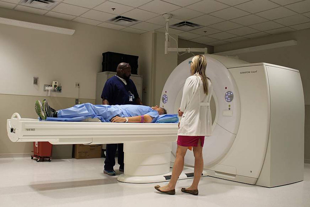
Image: A Spectral Photon Counting CT (SPCCT) scanner in use (Photo courtesy of US NIH).
The new technique was presented at the annual Radiological Society of North America (RSNA 2016) meeting by researchers from the Technical University Munich (Munich, Germany). The new protocols could enable clinicians to differentiate hemangioma and other liver abnormalities from Hepato-Cellular Carcinoma (HCC), for example, and spare patients with only small benign liver lesions, from undergoing unnecessary and expensive procedures.
The Results showed that by using SPCCT and an optimized contrast injection protocol, it was possible to provide contrast-enhanced images in a single CT scan, reducing radiation dose. The scan showed both arterial gadolinium distribution, and the portal-venous phase of iodine. The liver lesions, as well as the arterial and portal-venous contrast enhancement patterns were also visible in the scan. According to the researchers, the new technique eliminates mistaken registration of artifacts between acquisitions.
Daniela Muenzel, MD, Laboratory for Advanced Computed Tomography Imaging, Technical University of Munich, said, "This multi-phase visualization of the liver at one time point by a single CT scan exhibits perfect co-registration of the images in different phases, allowing for more accurate and quantitative subsequent voxel-by-voxel post processing and a significant reduction in radiation dose. By using two contrast agents and different uptake characteristics in liver lesions, we can classify cysts, hemangiomas, HCC and metastases in a single CT scan.
Related Links:
Technical University Munich


