Molecular Imaging Could Become the Standard for Prostate Cancer Tumor Staging
By MedImaging International staff writers
Posted on 28 Jun 2016
Researchers have shown that molecular imaging can help oncologists accurately detect and delineate prostate tumors during biopsies, and can also be used for pre-operative planning.Posted on 28 Jun 2016
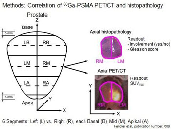
The researchers used Prostate-Specific Membrane Antigen (PSMA) Positron Emission Tomography/Computed Tomography (PET/CT) imaging and histopathology to evaluate prostate tumors of study participants. The researchers found that by using PET/CT imaging to evaluate Ga-68 PSMA tumor uptake, they were able to find all 67% of tissues found to be cancerous in a histological evaluation. PSMA is found on the surface of prostate cancer cells. The researchers used a combination of radioactive gallium-68 (Ga-68), and PSMA-HBED-CC a molecular compound to detect prostate cancer cells.

Image: These images compare axial histopathology and axial PET/CT in six segments of the prostate gland of a patient (Photo courtesy of Ludwig-Maximilians-Universitat of Munich).
The researchers presented the results at the annual meeting of the Society of Nuclear Medicine and Molecular Imaging (SNMMI 2016).
Wolfgang P. Fendler, MD, NM department, Ludwig-Maximilians Universitat Munchen (LMU; Munich, Germany), said, "PSMA shows significant over-expression on prostatic cancer cells and Ga-68 PSMA PET/CT demonstrates a high rate of detection in patients with recurrent, metastatic prostate cancer. However much less research has been conducted for the accuracy of PSMA imaging at the start of the disease. The results of our study indicate that Ga-68 PSMA PET/CT accurately identifies affected regions of the prostate and might thus present a promising tool for non-invasive tumor characterization and biopsy guidance."
Related Links:
Ludwig-Maximilians Universitat Munchen


