Narrow Band Imaging Can Reduce Bladder Tumor Recurrence
By MedImaging International staff writers
Posted on 09 Jun 2016
A new study concludes that narrow band imaging (NBI) technology can significantly reduce the risk of bladder tumor recurrence.Posted on 09 Jun 2016
Researchers at Harasanshin Hospital (Fukuoka, Japan), the University of Birmingham (United Kingdom), and other institutions conducted a randomized study in 965 patients with primary non–muscle-invasive bladder cancer following transurethral resection of bladder tumor (TURBT). The patients were randomized to NBI compared to white light (WL) guidance, the established imaging modality for TURBT. The primary outcome was 12-month bladder tumor recurrence rates.

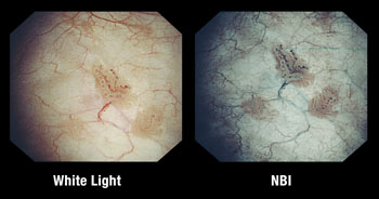
Image: A comparison of white light to narrow band imaging (Photo courtesy of Olympus Medical Systems).
The results showed WL-assisted TURBT recurrence rates of 27.1% and NBI-assisted TURBT recurrence rates of 25.4%. In patients at low risk for disease recurrence, recurrence rates at 12 months were significantly higher in the WL group (27.3%) than in the NBI group (5.6%). Although TURBT took longer on average with NBI plus WL compared with WL alone, lesions were significantly more often visible. The study was published on April 24, 2106, in European Urology.
“Narrow band imaging makes it easier to identify bladder tumors. It can detect small bladder tumors that might otherwise by overlooked by more conventional 'white light' cystoscopy,” said study co-author Richard Bryan, MD, PhD, of the University of Birmingham. “My colleague, Mike Wallace, and I immediately saw the potential of this technology. Now this potential has been confirmed by a large international randomized controlled trial, and the results can only be good news for bladder cancer patients worldwide.”
NBI technology, developed by Olympus Medical Systems (Tokyo, Japan), is a powerful optical enhancement technology that improves the visibility of blood vessels and other structures on the bladder mucosa by illuminating them with two specific wavelengths strongly absorbed by hemoglobin. The shorter wavelength (415 nm) only penetrates the top layer of the mucosa, and shows up as brown on the NBI image, The longer wavelength (540 nm) is absorbed by blood vessels located deeper within the mucosal layer, and appears cyan on the NBI image.
Related Links:
Harasanshin Hospital
University of Birmingham
Olympus Medical Systems


