Study Shows Safety and Efficacy of Imaging Technique for Neuroendocrine Tumors
By MedImaging International staff writers
Posted on 25 May 2016
The results of a new study have demonstrated the safety and efficacy of Ga-68 DOTATATE PET/CT scans, compared to In-111 pentetreotide scans, the current US imaging standard for the detection Neuroendocrine Tumors (NETS).Posted on 25 May 2016
The study showed that the use of Ga-68 DOTATATE imaging could significantly impact treatment management, resulting in no significant toxicity, a reduction in radiation exposure, and improved accuracy compared to the current standard in the US for the diagnosis and management of NETS. The US FDA has not yet approved the technique.

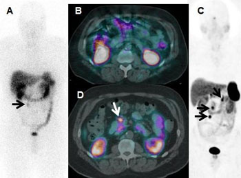
Image: The images demonstrate that Ga-68 DOTATATE PET/CT anterior 3D MIP and axial fused images could visualize metastases and change the surgical plan for resection (Photo courtesy of Ronald C. Walker, MD / Journal of Nuclear Medicine).
The study was performed by researchers at the Vanderbilt University Institute of Imaging Science (Nashville, TN, USA) and was published in the May 2016, issue of the Journal of Nuclear Medicine. The researchers enrolled 97 patients with known or suspected pulmonary or gastroenteropancreatic (GEP) NETS.
Corresponding author for the study, Ronald C. Walker, MD, professor of clinical radiology and radiological sciences, Vanderbilt University School of Medicine, said, "Our purpose was to evaluate the safety and efficacy of Ga-68 DOTATATE PET/CT compared to In-111 pentetreotide imaging for diagnosis, staging and re-staging of pulmonary and gastroenteropancreatic neuroendocrine tumors. Hopefully, our investigation will provide sufficient evidence on the safety and efficacy of Ga-68 DOTATATE to the U.S. FDA to allow approval. If so, then patients throughout the United States could soon have access to a higher-quality scan, allowing better patient management decisions while also lowering radiation exposure and shortening examination time."
Related Links:
Vanderbilt University Institute of Imaging Science


