Forensic Researchers Set Standards for X-Ray Identification
By MedImaging International staff writers
Posted on 12 Apr 2016
A new study establishes science-based standards for identifying human remains, based on X-rays of an individual's spine, upper leg, or the side of the skull.Posted on 12 Apr 2016
Researchers at North Carolina State University (NCSU; Raleigh, USA), Middle Tennessee State University (Murfreesboro, USA), and the University of South Florida (USF; Tampa, USA) compared ante-mortem and post-mortem lateral craniofacial X-rays in 41 cases, X-rays of the vertebral column in 100 cases, and X-rays of the proximal femur in a further 49 cases. The X-rays were then scored for number of concordant features, analyzed using classification decision trees, and evaluated using a receiver operating characteristic.

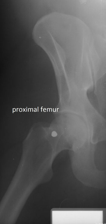
Image: Proximal femur concordant feature (Photo courtesy of Ann Ross/NCSU).
The researchers then used the data to develop specific standards for each skeletal region. They used additional, unmatched X-rays to test the accuracy of the standards in accurately identifying a body, and how likely were false-positive or false-negative results. The outcome showed a wide spectrum of consistencies; for example, two or more points of concordance are required in lateral cranial X-rays for a 97% probability of a correct identification, with a 10% misclassification rate. And just a single concordant feature is needed on cervical vertebrae for a 99% probability of correct identification, with a 7% misclassification rate.
And if there are one or more femoral head and neck concordant features, the probability of a correct identification is 94% and 97%, respectively. However, at the other end of the spectrum, four or more concordant features are required for a 98% probability of correct identification, and even there is a 40% misclassification rate. The study also established the minimum number of concordant areas needed to confirm positive identifications in the three standard radiographic views. The study was published on March 17, 2016, in in the American Journal of Forensic Medicine and Pathology.
“In the past, forensic experts have relied on a mixed bag of standards when comparing ante-mortem and post-mortem X-rays to establish a positive identification for a body, but previous research has shown that even experts can have trouble making accurate identifications," said lead author Professor of Anthropology Ann Ross, PhD, of NCSU. “We've created a set of standards that will allow for a consistent approach to identification that can be replicated, and that allows experts to determine probabilities for an identification.”
Related Links:
North Carolina State University
Middle Tennessee State University
University of South Florida


