3D Noninvasive Virtual Autopsies Link Forensics and Radiology
By MedImaging International staff writers
Posted on 05 Apr 2016
Virtual autopsies that create 3D Computed Tomography (CT) models of the body, while leaving it intact, represent the future forensic medicine.Posted on 05 Apr 2016
The new method does not interfere with forensic evidence, and 3D models can easily be shared for obtaining a second opinion. The cost of virtual autopsies is still a barrier, but they are expected to come down with the introduction of new technology, and increased acceptance of the new method.

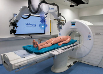
Image: The robotic Virtobot system performs various tasks together with a CT scanner to produce high-resolution 3D surface documentation, and enable CT-guided postmortem tissue sampling. (Photo courtesy of RSNA).
Virtual autopsies were pioneered by Dr. Thali, Institute of Forensic Medicine at the University of Zurich (Zurich, Switzerland), who also co-founded The Virtopsy Project. The new method is already standard procedure in Switzerland, and is gradually being accepted around the world.
The Virtobot, a robotic system used for virtual autopsies, works with a CT scanner to create automated, high resolution 3D surface images, and performs CT-guided post-mortem tissue sampling for documentation of an injury. Bite marks, for example, can be modeled in 3D for comparison with the dental records of a suspect for use as evidence.
Virtual autopsies also take less time because imaging can be performed quickly. They are observer-independent enabling objective data archiving, and can be used in situations where conventional autopsies are not possible for religious or other reasons.
Dr. Thali, said, “With virtual autopsy, imaging becomes the gold standard in the future examination of forensic evidence. At the moment, we cannot see everything with imaging, but judging by the (technology) on display at RSNA 2015, I think the direction is absolutely clear. Our customer (the court system) often has no real knowledge of the body’s internal structures, so having 3D visualization is a good tool to show what really happened to the body.”
Related Links:
Institute of Forensic Medicine, University of Zurich


