Low-Dose Radiography Effective for Limb Measurement
By MedImaging International staff writers
Posted on 28 Feb 2016
Low-dose biplanar radiography may perform as well as conventional computed tomography (CT) scans in assessing limb length, according to a new study.Posted on 28 Feb 2016
Researchers at the Hospital for Special Surgery (HSS; New York, NY, USA) conducted a study to determine the accuracy and reliability of the EOS Imaging System as compared with a CT scanogram for the measurement of leg length. To do so, 0.8 mm diameter Tantalum beads were inserted into the cortex on both the medial and lateral sides of 10 skeletally immature lamb femurs. CT scanogram and EOS imaging were then obtained, and measurements of total length and distance between bead pairs were recorded on both anteroposterior and lateral views.

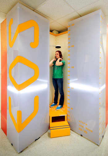
Image: The EOS imaging system (Photo courtesy of EOS Imaging).
The results revealed that the biplanar EOS measurements, as evaluated by two orthopedic surgeons, showed near-perfect correlation to those of CT scanogram, and intrarater and interrater reliability were excellent for all measurements with both EOS and CT scanogram. According to the researchers, the combination of EOS imaging and tantalum bead implantation may be an effective way to evaluate physeal growth following procedures such as epiphysiodesis and physeal bar resection. The study was published in the January 2016 issue of the Journal of Pediatric Orthopaedics.
“CT or conventional scanograms are the current gold standard for measuring limb-length discrepancy. However, the use of low-dose EOS is being used more frequently in pediatric orthopedics,” said senior author Emily Dodwell, MD, MPH. “The greatest benefit of using these tantalum markers with EOS may be in monitoring growth following injury or surgery to the growth plate. Further investigations are necessary to determine whether this measurement technique will be of significant clinical utility.”
The EOS Imaging System, a product of EOS Imaging (Paris, France) is a low-dose, system that provides three-dimensional (3D) images of patients in natural standing positions using perpendicular X-ray beams collimated in two very thin, horizontal, fan-shaped beams. Two variable gain detectors provide an extremely high contrast digital radiograph using a significantly lower radiation dose than a general radiography X-ray, thus enabling clinicians to make a more informed diagnosis and create individualized treatment plans for children with musculoskeletal disorders.
Related Links:
Hospital for Special Surgery
EOS Imaging


