3D Printing Supports Accurate Radiation Therapy Delivery
By MedImaging International staff writers
Posted on 08 Jul 2015
A new study demonstrates how an inexpensive three dimensional (3-D) printer can be used to manufacture a patient-specific bolus for external beam therapy.Posted on 08 Jul 2015
Researchers at Stony Brook Medicine (NY, USA) conducted a project to design and print a bolus using a treatment planning system and an inexpensive (USD 3,000) 3-D printer. The bolus is a device used in radiation therapy (RT) placed directly on the patient’s skin, and is intended to shape the desired RT dose to surface anatomy, thus conforming and containing the planning target volume (PTV) dose while delivering minimal radiation to adjacent underlying critical structures and normal tissues.
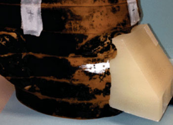
Image: Example of bolus printed to conform to the nose of a phantom (Photo courtesy of Stony Brook Medicine).
The researchers began the design process using a phantom as the test subject. After a computerized tomography (CT) scan was acquired, the data was exported to the Varian (Palo Alto, CA, USA) Eclipse treatment planning system. Once a satisfactory bolus design was determined, the structure set was exported to 3DSlicer, a 3-D modeling software that is maintained as open source. The stereolithography (STL) files were interpreted by printer software, and instructions were sent to an Airwolf (Costa Mesa, CA, USA) 3-D printer.
The researchers tested different materials—including acrylonitrile butadiene styrene and polylactic acid—as the substrate. Dose plane comparisons were conducted for each material using the phantom model and photographic film to verify accurate treatment planning. They were also able to verify accurate treatment planning using gamma analysis, and found that with gamma criteria of 5% dose difference and 3 mm distance-to-agreement (DTA) leeway, they achieved 95% points passing. The study was published in the May-June 2015 issue of the Journal of Applied Clinical Medical Physics (JACMP).
“We are confident that we can accurately model this printing material in our treatment planning system for all energies in photon and electron beams,” concluded lead author Sarah Burleso, PhD, and colleagues of the department of radiation oncology. “In the event the patient cannot have bolus materials placed on their skin for molding, we can print a 3D positive mold of the patient’s treatment area, molding the bolus to the replica instead. This process of printing our own bolus streamlines patient care, minimizes patient involvement, and maintains quality treatments.”
Related Links:
Stony Brook Medicine
Varian
3DSlicer




X-Ray Generator
Advantage Plus Generators

Medical Radiographic X-Ray Machine
TR30N HF

Ultrasound Table
Women’s Ultrasound EA Table
New

Radiology Monitor
Barco MDNC-12130 Nio Fusion 12MP Radiology Monitor