New Study Investigates Use of Multi-Trauma Computed Tomography Examinations
By MedImaging International staff writers
Posted on 04 Mar 2015
A study examined ways the introducing Whole-body Computed Tomography (WBCT) affected the emergency care workflow.Posted on 04 Mar 2015
Researchers compared Computed Tomography (CT) workflow of 120 consecutive trauma patients in the hospital before the introduction of the WBCT and another 120 trauma patients afterwards. The Researchers compared the number and type of CT exams carried out, the number of radiography exams, Focused Assessment with Sonography (FAST) for trauma, and any additional CT exams made. The time it took to complete trauma imaging exams was also measured.

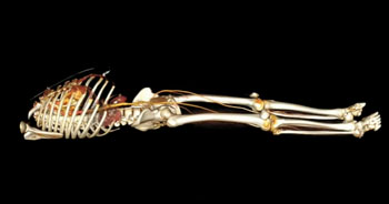
Image: Whole body CT scan with contrast media (Photo courtesy of Atelier KMR).
The study was carried out by researchers at the University Hospital Zurich, (Zurich, Switzerland) and published online in the February 11, 2015, issue of the British Journal of Radiology.
The results showed that patients undergoing imaging after the WBCT was in use, were significantly more likely to undergo head, neck, chest, and abdomen CT exams (p < 0.001), compared to the non-WBCT group. Patients in the WBCT group underwent significantly less radiographic exams of the chest, cervical spine, pelvis, and FAST exams (p < 0.001). Radiographic examinations of the upper extremities (p = 0.56) and lower extremities (p = 0.30) were not significantly different. Patients in the WBCT group were exposed to significantly higher effective doses of radiation (29.5 mSv) than those in the non-WBCT group (15.9 mSv; p < 0.001).
However, fewer additional CT exams were necessary to complete the work-up in the WBCT group (p < 0.001). The complete trauma-related imaging workflow took significantly less time for patients in the WBCT group (12 min) compared to those in the non-WBCT group (75 min; p < 0.001).
The researchers concluded that for trauma patients, the use of WBCT in the initial work-up resulted in higher radiation doses, while on the other hand less additional CT exams were needed, and the trauma-related imaging workflow took significantly less time. The radiation dose from WBCT for trauma patients was 29.5 mSv.
Related Links:
University Hospital Zurich


