Device Designed to Lessen Mammography Discomfort and Pain
By MedImaging International staff writers
Posted on 09 Dec 2014
Researchers have developed a new device that may result in more comfortable mammography for women. Posted on 09 Dec 2014
According to a recent study, standardizing the pressure applied in mammography screening would lessen the pain and discomfort associated with breast compression without sacrificing image quality.

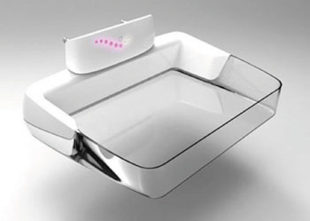
Image: A retrofit paddle for an existing mammography machine that has been in use in the Academic Medical Center Amsterdam since December 2013 (Photo courtesy of RSNA).
Compression of the breast is required in mammography procedures to optimize image quality and minimize absorbed radiation dose. However, mechanical compression of the breast in mammography frequently causes discomfort and pain and deters some women from mammography screening. An additional difficulty tied to compression is the variation that occurs when the technologist adjusts compression force to breast size, composition, skin tautness and pain tolerance. Over-compression, or unreasonably high pressures during compression, is common in certain European countries, particularly for women with small breasts. Over-compression occurs less frequently in the United States, where under-compression, or extremely low applied pressure, is more common.
“This means that the breast may be almost not compressed at all, which increases the risks of image quality degradation and extra radiation dose,” said Woutjan Branderhorst, PhD, researcher in the department of biomedical engineering and physics at the Academic Medical Center (Amsterdam, The Netherlands). The study’s findings were presented December 2014 at the annual meeting of the Radiological Society of North America (RSNA), held in Chicago (IL, USA).
Overall, adjustments in force can lead to substantial variation in the amount of pressure applied to the breast, ranging from less than 3 kPa to greater than 30 kPa. Dr. Branderhorst and colleagues theorized that a compression protocol based on pressure rather than force would reduce the pain and variability associated with the current force-based compression protocol. Force is the total impact of one object on another, whereas pressure is the ratio of force to the area over which it is applied.
The researchers developed a device that displays the average pressure during compression and studied its effects in a double-blinded, randomized control trial on 433 asymptomatic women scheduled for screening mammography. Three of the four compressions for each participant were standardized to a target force of 14 dekanewtons (daN). One randomly assigned compression was standardized to a target pressure of 10 kPa.
Participants scored pain on a numeric rating scale, and three experienced breast screening radiologists indicated which images required a retake. The 10 kPa pressure did not compromise radiation dose or image quality, and, on average, the women reported it to be less painful than the 14 daN force.
The study’s findings are potentially important, according to Dr. Branderhorst. There are an estimated 39 million mammography scans performed every year in the United States alone, which translates into more than 156 million compressions. Pressure standardization could help avoid a large amount of unnecessary pain and optimize radiation dose without adversely affecting image quality or the proportion of required retakes. “Standardizing the applied pressure would reduce both over- and under-compression and lead to a more reproducible imaging procedure with less pain,” Dr. Branderhorst said.
The device that displays average pressure is easily added to existing mammography systems, according to Dr. Branderhorst. “Essentially, what is needed is the measurement of the contact area with the breast, which then is combined with the measured applied force to determine the average pressure in the breast,” he said. “A relatively small upgrade of the compression paddle is sufficient.”
Additional studies, according to the investigators, will be needed to determine if the 10 kPa pressure is the optimal target. The researchers are also working on new methods to help mammography technologists enhance compression through more optimized positioning of the breast.
Related Links:
Amsterdam Academic Medical Center


