Wireless X-Ray Image Detectors Optimized for Detailed Imaging at Low Dose Rates
By MedImaging International staff writers
Posted on 12 Mar 2013
New dual-band X-ray detectors are licensed for use in most markets worldwide. They can transmit image data in real-time across a very consistent wireless link. Posted on 12 Mar 2013
A range of digital X-ray image detectors are portable, effective, fast, usable worldwide, and capable of generating highly detailed images at low dose rates. These features are among the X-ray imaging system components that Varian Medical Systems (Palo Alto, CA, USA) presented at the annual meeting of the European Congress of Radiology (ECR) in Vienna, Austria, from March 7-11, 2013.

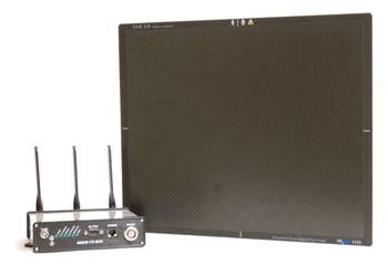
Image: The PaxScan 4336W wireless image detector (Photo courtesy of Varian Medical Systems).
“We will be showing our new PaxScan 4336W wireless image detector, which was engineered to be dose efficient and capable of providing excellent image quality, even at low doses,” stated Steve Kimmel, vice president of sales and marketing for Varian X-ray products. “These detectors were designed for use in systems that can provide clinicians with the details they need to make accurate medical decisions while keeping X-ray dose to a minimum.”
“Even with the panel inside a cassette tray, or down below the X-ray table, the transmission rates stay consistently high,” stated Rick Colbeth, vice president of engineering for Varian imaging products. “Quality images can be generated very quickly.”
The wireless panel also includes a built-in diagnostic program that provides a way to continuously monitor the “health” of the unit for faster, easier servicing. In addition to the new wireless panel, Varian will also present other X-ray imaging products and technologies.
The PaxScan 4343CB is a sophisticated radiography and fluoroscopy panel that provides high quality X-ray images as well as excellent low-dose fluoroscopic images, showing motion at up to 60 frames per second. “This panel has one of the smallest pixel sizes available, which yields a higher quality image in terms of resolution,” Mr. Colbeth said.
The PaxScan 3024M is a full-field digital mammography detector designed for rapid image acquisition and tomosynthesis, a method of using X-rays to create a three-dimensional [3D] image of the breast. “This technology enables customers to extend the capabilities and efficiencies of digital mammography into emerging markets and mobile screening applications,” noted Mr. Kimmel.
The PaxPower M-1500 X-ray tube was developed specifically for advanced mammography applications such as tomosynthesis and CT scanning. Its space-efficient design enables high quality imaging in the chest wall region, as well as the power to enable sophisticated slit scanning and photon counting approaches that can yield higher contrast when imaging large or dense areas.
Varian’s InfiMed image processing software and workstations offer cutting-edge imaging algorithms for processing data from radiographic flat panel image detectors, dynamic detectors, and legacy image intensifier systems. “Our imaging workstations enable customers to add digital dynamic or radiographic productivity to their portfolios, reducing time to market and enhancing their competitive position,” Mr. Kimmel remarked.
Related Links:
Varian Medical Systems


