Portable X-Ray Source Brings Sci-Fi Closer to Reality
By MedImaging International staff writers
Posted on 23 Jan 2013
Tricorders, the Star Trek television version of futuristic hand-held medical scanners, are closer to becoming a reality now that engineers have designed a compact source of X-ray and other types of radiation. The radiation source, the size of a stick of gum, could be used to create portable and cost-effective X-ray scanners, as well as used in other critical applications. Posted on 23 Jan 2013
“Currently, X-ray machines are huge and require tremendous amounts of electricity,” said Dr. Scott Kovaleski, associate professor of electrical and computer engineering at the University of Missouri (MU; Columbia, USA). “In approximately three years, we could have a prototype hand-held X-ray scanner using our invention. The cell-phone-sized device could improve medical services in remote and impoverished regions and reduce healthcare expenses everywhere.”

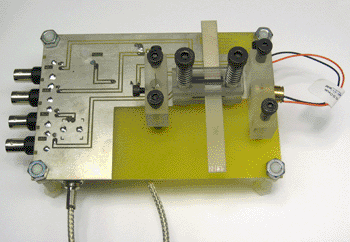
Image: The compact radiation source developed by Kovaleski's team (Photo courtesy of Peter Norgard, University of Missouri Journalists).
Dr. Kovaleski recommends other uses for the device. In dentists’ offices, the little X-ray generators could be used to take images from the inside of the mouth shooting the X-rays outward, reducing radiation exposure to the rest of the patients’ heads. At ports and border crossings, portable scanners could search cargoes for contraband, which would both reduce costs and improve security. Interplanetary probes, such as the Curiosity rover, could be equipped with the compact sensors, which otherwise would require too much energy.
The accelerator developed by Dr. Kovaleski’s team could be used to create other types of radiation in addition to X-rays. For example, the development could replace the radioactive materials, called radioisotopes, used in drilling for oil as well as other industrial and scientific operations. The instrument could replace radioisotopes with a safer source of radiation that could be turned off in case of emergency. “Our device is perfectly harmless until energized, and even then it causes relatively low exposures to radiation,” said Dr. Kovaleski. “We have never really had the ability to design devices around a radioisotope with an on-off switch. The potential for innovation is very exciting.”
The device uses a crystal to produce more than 100,000 volts of electricity from only 10 volts of electrical input with low power consumption. Having such a low need for power could allow the crystal to be fueled by batteries. The crystal, made from a substance called lithium niobate, uses the piezoelectric effect to amplify the input voltage.
The study’s findings were published January 2013 the journal IEEE Transaction on Plasma Science.
Related Links:
University of Missouri


