Appendicitis Study Shows Benefits of CT Scans
By MedImaging staff writers
Posted on 25 Mar 2008
Posted on 25 Mar 2008

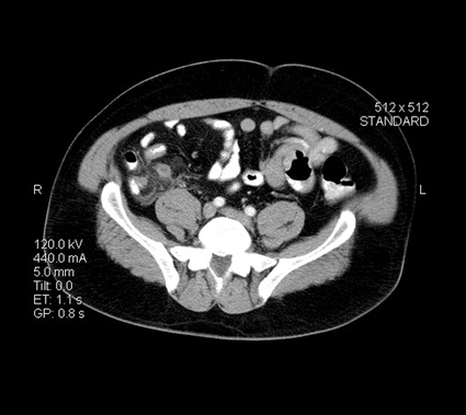
Image: Axial cross-sectional CT image through the upper pelvis showing acute appendicitis, with an enlarged appendix and streaky inflammatory changes in the surrounding mesenteric fat (Photo courtesy of Living Art Enterprises).
The Medical Imaging & Technology Alliance (MITA), a division of the U.S. National Electrical Manufacturers Association (NEMA; Roslyn, VA, USA), recently commented on the study performed by researchers from the University of California, Los Angeles (UCLA; Los Angeles, CA, USA).
"Medical imaging has become integral to best practices across so many disease states and certainly plays a critical role in providing high quality patient care,” said Dr. Andrew Whitman, vice president, MITA. "MITA applauds the work of Dr. Steven Raman and his team at UCLA. Their research findings remind us that it's critical that patients have access to innovative medical imaging technology to help fight serious illnesses.”
Dr. Whitman also reported that this UCLA study is one of many reinforcing the value of abdominal CT scans. For example, a June 2007 article in the journal The American Surgeon highlighted research showing that use of abdominal CT scanning in women of reproductive age can reduce the rate of unnecessary appendectomies by 58%. Furthermore, that study demonstrated that use of abdominal CT scanning does not increase patient and insurance company costs, but instead, provides a net savings of US$1,412 per patient when weighed against the cost of negative appendectomy--not only for women, but for male patients as well.
"Now more than ever it's crucial that patients and policymakers alike look to studies like these as a reminder of why and how medical imaging improves patient health outcomes and reduces overall healthcare costs,” Dr. Whitman said.
Related Links:
National Electrical Manufacturers Association
University of California, Los Angeles


