Human Prostate Cancer Study Evaluates New Imaging Biomarker
By MedImaging International staff writers
Posted on 26 May 2015
The results of the first human prostate cancer study intended to evaluate a 2nd generation fluorine-18 labeled small-molecule PSMA inhibitor, have been published in the April 28, 2015, issue of the Journal of Molecular Imaging and Biology.Posted on 26 May 2015
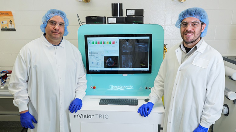
The results of the evaluation demonstrated the initial safety, bio-distribution, and dosimetry results with the [18F]DCFPyL imaging biomarker, which was developed at Johns Hopkins University (Baltimore, MD, USA).
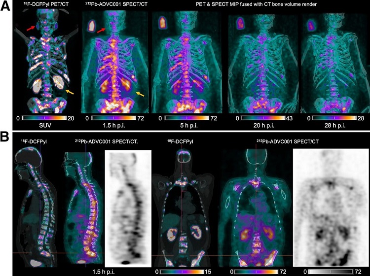
A large number of prostate cancer patients remain with metastases after treatment, and require further imaging to detect lesions, for therapeutic monitoring, and restaging. Existing imaging techniques are not sensitive or specific enough to detect these lesions. The results of the study show that the [18F]DCFPyL radiotracer was safe, and compared to the first generation radiotracer was able to parallel the expected uptake with much improved visual conspicuity of suspected lesions.
The [18F]DCFPyL biomarker is a PET agent that attaches to the Prostate-Specific Membrane Antigen (PSMA) and can subsequently be measured using a Positron Emission Tomography (PET) scan. The biomarker produced images that providing clearer images than the first generation agent and resulted in 50% less radiation dose in the most sensitive organs.
Dr. Jason Lewis, President of the World Molecular Imaging Society (WMIS) said, “The basis of more accurate, molecularly-informed classification of disease is the premise of precision medicine and specific molecular imaging biomarkers are the keys to determine how we classify diseases, how we select therapy, how we monitor therapy, and ultimately how we make treatments more accurate for each individual for better patient outcomes. We commend the team at Johns Hopkins for developing a more sensitive and accurate PSMA.”
Related Links:
Johns Hopkins University
WMIS

Gold Member
Solid State Kv/Dose Multi-Sensor
AGMS-DM+

PACS Workstation
CHILI Web Viewer

New
Illuminator
Trimline Basic

DR Flat Panel Detector
1500L