SPECT/CT Technology Offers High Resolution and Quantitative Imaging
By MedImaging International staff writers
Posted on 16 Jul 2014
New imaging technology incorporates single-photon emission computed tomography (SPECT) and computed tomography (CT) during image reconstruction, combining SPECT’s high sensitivity with high resolution, and for the first time, quantitative images.Posted on 16 Jul 2014
The University of Minnesota Medical Center-Fairview Health Services (Minneapolis, MN, USA) recently became the first US healthcare facility to install the Symbia Intevo xSPECT system, developed by Siemens Healthcare (Erlangen, Germany).
.")
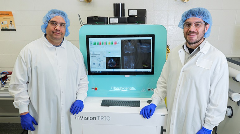
Image: The Symbia Intevo xSPECT SPECT/CT system (Photo courtesy of Siemens Healtcare).
“The Symbia Intevo’s ability to truly merge SPECT and CT data provides our physicians with invaluable additional diagnostic information, helping us to differentiate cancer from other forms of disease,” said Jerry Froelich, MD, director of nuclear medicine and molecular imaging at the University of Minnesota. “For example, with the Intevo, we are better able to identify metastatic change within an area of degenerative change, such as the spine of older patients. The supporting information of the system’s xSPECT image enables us to make these kinds of objective interpretations of disease in cases where subjective interpretations had only been possible previously.”
The Symbia Intevo xSPECT system reconstructs both the SPECT and CT portions of the image into a much higher frame of reference than previous systems. The result is precise, accurate alignment of SPECT and CT that facilitates the extraction and deep integration of medically relevant information. This ability is also the basis for differentiating tissue boundaries in bone imaging. With xSPECT Bone, physicians can potentially provide additional support for identifying and differentiating between cancerous lesions and degenerative disorders. In addition, single-step reading with integrated xSPECT Bone images may reduce physician time to read and report.
xSPECT Quant delivers precise alignment of SPECT and CT, providing physicians with essential volumetric information from the CT scan. This information enables accurate and consistent quantitative assessment—a numerical indication of a tumor’s level of metabolic activity. Effective quantitative evaluation enables the physician to assess whether a patient’s course of treatment has regressed, stabilized, or grown.
Related Links:
University of Minnesota Medical Center
Siemens Healthcare

Gold Member
Solid State Kv/Dose Multi-Sensor
AGMS-DM+

New
Ultrasound Table
Powered Ultrasound Table-Flat Top

New
Ultrasound Table
Ergonomic Advantage (EA) Line

Brachytherapy Planning System
Oncentra Brachy