'Dual-Mode' Tracer Enables Surgeons to See and Hear Prostate Cancer
Posted on 27 Aug 2025
Prostate cancer is one of the most common cancers in men, with about one in eight Canadian men developing the disease during their lifetime; one in 30 will die from it. Treatment often requires balancing the removal of tumors with the preservation of critical structures such as nerves, the seminal vesicle, bowel, and bladder. Surgeons need more precise tools to detect and remove cancer while reducing the risks of recurrence and surgical complications. Now, a new tracer could enable surgeons to see and hear prostate cancer.
Researchers at the University of British Columbia (Vancouver, Canada) have developed a new “dual-mode” tracer designed to help image, plan, and guide prostate cancer procedures. The tracer uses a single molecule labeled with Fluorine-18, a common isotope in PET scans, enabling both fluorescence-guided and radio-guided surgery. By combining 18F-organotrifluoroborates with fluorescein, the tracer offers both high-resolution visual guidance and radioactive detection through handheld Geiger counter probes.

The tracer works by targeting PSMA, a protein highly expressed on prostate cancer cells. It demonstrates high tumor uptake for PET imaging and strong optical brightness in fluorescent mode without requiring special visual equipment. Unlike other approaches that require separate injections of different tracers, this method allows clinicians to both see and “hear” radiation signals from cancerous tissue, including lymph nodes or metastases.
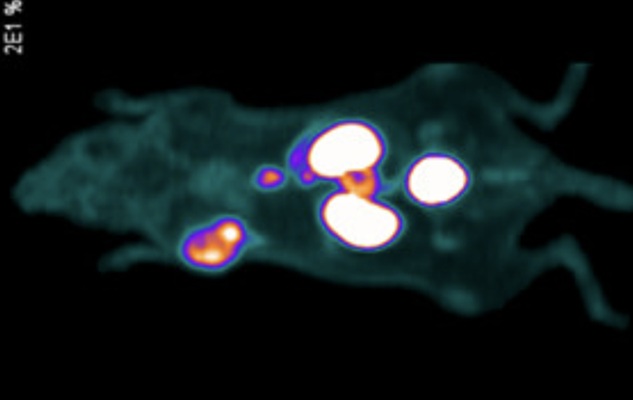
In preclinical testing, UBC scientists and collaborators at BC Cancer trialed the tracer on mice implanted with human tumors. The findings, published in the Journal of Medicinal Chemistry, showed high uptake and strong signals in cancerous tissues compared to benign samples, demonstrating feasibility for both diagnostic imaging and surgical guidance.
The dual-mode tracer could reduce unnecessary biopsies, improve the precision of surgery, and expand access to advanced imaging in smaller hospitals with standard equipment. Researchers plan Good Manufacturing Practices assessments, toxicity testing, and validation studies to prepare for human trials. They also suggest potential applications beyond prostate cancer, including ovarian and laryngeal cancers.
“The implementation of dual-mode fluorescent-PET tracers in the surgical field is an exciting new approach to maximize benefit and minimize harm associated with more extended lymph node removal as well as to decrease the rate of positive surgical margins of a radical prostatectomy,” said Dr. Larry Goldenberg, associate director of development and supportive care at the Vancouver Prostate Center and professor of urologic sciences at UBC.
Related Links:
BC Cancer Research
University of British Columbia


