AI Algorithms Accurately Predict Tumor Location and Size from Medical Images
Posted on 02 Dec 2024
Cancer patients often have numerous lesions, or pathological changes caused by tumor growth, and capturing all of them is crucial to obtaining a comprehensive view of their condition. Imaging plays a vital role in diagnosing cancer, as accurately determining the location, size, and type of tumors is essential for selecting the appropriate treatment. Two key imaging techniques used are positron emission tomography (PET) and computed tomography (CT). PET uses radionuclides to visualize metabolic processes in the body, as the metabolic activity of malignant tumors is significantly higher than that of benign tissue. Fluorine-18-deoxyglucose (FDG), a radioactively labeled glucose, is commonly used for this purpose. In contrast, CT scans the body layer by layer with an X-ray tube to visualize anatomical structures and locate tumors. Doctors often manually mark tumor sizes on 2D slice images, a process that is both time-consuming and labor-intensive.
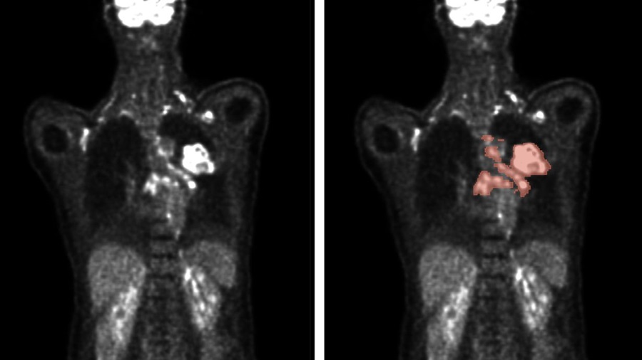
Artificial intelligence (AI) has shown great promise in enhancing the analysis of medical images. Deep learning algorithms, for example, can automatically identify tumor locations and sizes. By automating this process, significant time savings can be achieved, and results can be more consistent and accurate. The seven best teams participating in AutoPET, an international competition in medical image analysis, have now reported in the journal Nature Machine Intelligence on how algorithms can detect tumor lesions in PET and CT. Researchers from the Karlsruhe Institute of Technology (KIT, Karlsruhe, Germany) participated in the 2022 AutoPET competition and secured fifth place out of 27 teams, with 359 participants from around the globe. The competition tasked teams with automatically segmenting metabolically active tumor lesions visualized in whole-body PET/CT scans.

The teams used a large annotated PET/CT dataset for training their algorithms, all of which were based on deep learning techniques. This form of machine learning utilizes multi-layered artificial neural networks to detect complex patterns and correlations within large datasets. The results, now published in Nature Machine Intelligence, show that combining the top-performing algorithms into an ensemble approach outperforms individual models in detecting tumor lesions with high efficiency and precision. The researchers note that further refinement of these algorithms is necessary to improve their resilience to external variables, so they can be effectively implemented in routine clinical settings. The ultimate goal is to fully automate the analysis of PET and CT medical images in the near future.
“While the performance of the algorithms in image data evaluation partly depends indeed on the quantity and quality of the data, the algorithm design is another crucial factor, for example with regard to the decisions made in the post-processing of the predicted segmentation,” explained KIT researcher Rainer Stiefelhagen.


