PET/CT Imaging Using New Tracing Agent Could Become ‘Gold Standard’ Test for Prostate Cancer Detection
Posted on 03 Jul 2024
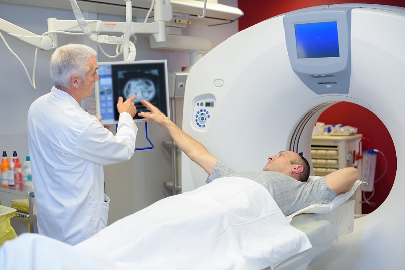
The diagnosis and management of intermediate-risk and high-risk prostate cancer increasingly benefit from advancements in diagnostic imaging. Present guidelines advocate for the use of multiparametric magnetic resonance imaging (MRI) to aid in the diagnosis and locoregional staging of prostate cancer prior to radical prostatectomy. Now, a new study indicates that positron emission tomography (PET)/computed tomography (CT) imaging, utilizing a novel tracer, is more effective at determining the extent of prostate cancer in intermediate and high-risk cases compared to traditional MRI. This method involves the administration of a new radioactive tracer specific to prostate tumors, 18F-PSMA-1007, followed by tracking with PET and CT technologies. Earlier attempts with other tracers in PET/CT scans have not shown similar effectiveness.
In research published this week in the journal JAMA Oncology, investigators at the University of Alberta (Edmonton, AB, Canada) conducted both PET/CT and MRI scans on 134 men scheduled for radical prostatectomy—the surgical removal of the prostate gland and surrounding tissue, including lymph nodes—within a two-week interval. The accuracy of these tests in predicting tumor size and location was then verified against the actual tumors identified during surgery. Results demonstrated that the new imaging test accurately located and delineated tumor margins in 45% of cases, nearly doubling the accuracy rate of MRI at 28%. This precision is critical for treatment planning; for instance, if cancer extends beyond the prostate, surgeons usually adjust their surgical margins to ensure no cancerous tissue is left behind. Similarly, radiation oncologists intensify radiation at the cancer's core for enhanced control when treating with radiation. This imaging test can improve the precision in targeting where treatments should be applied.

Although the test exposes patients to a minor amount of radiation, there were no adverse reactions reported in the study. The encouraging outcomes have led to the initiation of another clinical trial to explore whether the PET/CT scan can direct ablation techniques, which involve the destruction of cancer cells in the prostate using various forms of energy. The researchers believe that the PET/CT scan with this new tracer will set a new standard for detecting prostate cancer and expect it to eventually replace the need for additional CT and bone scans which are currently necessary for prostate cancer patients. This would reduce hospital visits, decrease waiting times for results, and lower radiation exposure for patients, although further research is required to confirm these benefits.
Related Links:
University of Alberta


