New Imaging Method Could Accurately Identify Serious Lung Infections in Real Time
Posted on 16 Aug 2023
Radiologists presently rely on chest X-rays to confirm the diagnosis of pneumonia and similar lung infections. However, X-rays fall short of specifying the nature of the infection as it does not confirm whether it's bacterial, viral, or fungal. To identify the exact type of infection, pathologists need to culture a sample from the lung, obtained through an invasive procedure known as bronchoscopy. This process usually takes 2-3 days. For patients in critical condition, especially those with conditions like chronic obstructive pulmonary disease (COPD) and infectious pneumonia, waiting isn't always an option.
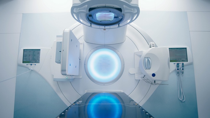
Now, researchers at the University of Cincinnati (Cincinnati, OH, USA) are developing an imaging technique that can identify specific lung infections in real time, thereby expediting treatment for critically ill patients who are most at risk. Supported by a five-year USD 3 million grant from the National Heart, Lung, and Blood Institute (NHLBI), the researchers will focus on designing and testing the efficacy of different types of injectable probes or metallic contrast agents. Once injected, these agents would accumulate at the infection site and become detectable in real time using a PET scan.
")
One key advantage of this new imaging method is the quick development process of the contrast agent that eliminates the need for elaborate processing or preparation time as compared to the time-consuming and complicated development of contrast agents. Streamlining this process will not only cut down lab preparation time but also pave the way for its broader clinical adoption. The researchers are currently testing the method in animal models, focusing on bacterial and viral pneumonia associated with COPD. However, the potential of this imaging method extends to other types of infections such as fungal infections or conditions like cystic fibrosis. Post-treatment imaging can provide insights into a patient's response to medications, like antibiotics, and even guide the choice of the most effective antibiotic by identifying the responsible pathogens.
“Our solution is to use imaging to identify what is causing the pneumatic episode within hours, to hasten a treatment plan,” said University of Cincinnati researcher Nalinikanth Kotagiri who is leading the study.
Related Links:
University of Cincinnati