Reconstructed MRI Images Guide Radiotherapy Treatment
By MedImaging International staff writers
Posted on 07 Jul 2021
An artificial intelligence (AI) algorithm that reconstructs magnetic resonance imaging (MRI) images of moving tumors in seconds could optimize radiotherapy (RT) treatment, according to a new study.Posted on 07 Jul 2021
Developed at The Institute of Cancer Research (ICR; London, United Kingdom), The Royal Marsden NHS Foundation Trust (London, United Kingdom), the German Cancer Research Center (DKFZ; Heidelberg, Germany), and other institutions, magnetic resonance guided radiotherapy (MRgRT) is based on a deep radial convolutional neural network (Dracula) algorithm that replicates four-dimensional (4D) MRI images, including tumors and healthy organs, from low quality images containing artifacts.
")
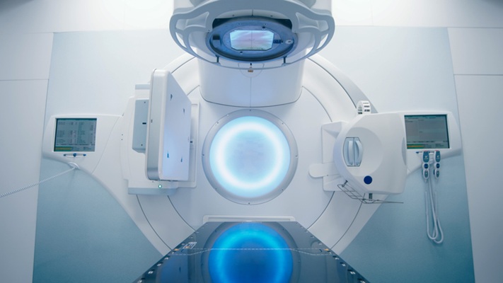
Image: The MRgRT treatment room at the ICR (Photo courtesy of ICR)
By using data on the spatial dimensions and respiratory phases of a patient's tumor and surrounding organs, Dracula can produce 4D MRI and mid-position images from scans previously considered unacceptable for MRgRT. Clinically, the MRI images reconstructed by Dracula can be used to guide a linear accelerator (Linac), thus serving as a novel platform to locate and deliver precisely targeted RT doses to tumors, minimizing the risk of affecting healthy organs. The study was published in the June 2021 issue of Radiotherapy and Oncology.
“Using an AI to rapidly reconstruct 4D MRI images of a cancer patient's anatomy lets us accurately determine the location of tumors and characterize their motion right before RT treatment,” said senior author Andreas Wetscherek, PhD, of the ICR. “It would be especially useful for cancers where we need to avoid the healthy organs around the tumor, such as pancreatic cancer, which currently limits the delivery of high doses of radiation to the tumor.”
To treat patients with tumors that move as they breathe, 4D MRI images are needed to show the 3D volume of the tumor and surrounding organs at different time points during breathing, known as a respiratory phase. By combining respiratory phases, the midposition (the average position of the tumor) is calculated, helping to accurately plan MRgRT. An alternative is to have a patient hold their breath, but this can be strenuous for the patient and makes treatment longer and more difficult.
Related Links:
The Institute of Cancer Research
The Royal Marsden NHS Foundation Trust
German Cancer Research Center

Medical Radiographic X-Ray Machine
TR30N HF

Diagnostic Ultrasound System
DC-80A

Half Apron
Demi

Portable X-ray Unit
AJEX140H