Radioactive Cement Safer for Spinal Tumor Treatment
By MedImaging International staff writers
Posted on 01 Mar 2021
Injecting brachytherapy cement into bone is a safer alternative to conventional radiation therapy (RT) for spinal tumors, according to a new study.Posted on 01 Mar 2021
Developed by researchers at the University of California Irvine (UCI, USA) Spine-Rad brachytherapy cement includes radioactive isotopes that are dispersed evenly, so that radiologists don’t need to measure the total amount of radioactivity; the dose delivered to the tumor is independent of the volume of cement and the amount injected. To validate the function of the cement, the researchers conducted animal and computational studies to evaluate the short-term efficacy, safety, migration of radioactivity into blood, urine, or feces; and the radiation dose from phosphorus-32 (P-32) emissions to the spinal cord and soft tissues.

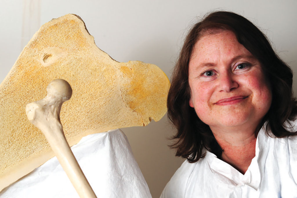
Image: Professor Joyce Keyak, co-developer of Spine-Rad brachytherapy cement (Photo courtesy of UCI)
The results showed that at 17 weeks post-injection, physical examinations were all normal, and no activity was detected in blood, urine or feces. The researchers found no evidence of the P-32 isotope in the circulating blood, no changes in blood work related to radioactivity, no neurological deficits, and that radiation dose rates outside the injection site were minimal. The study was presented at the annual meeting of the Orthopaedic Research Society, held virtually during February 2021.
“Currently, multiple sessions of external beam radiation are used to treat cancer that has spread to the spine. But with the brachytherapy bone cement, a single injection can provide an equivalent, targeted tumor treatment with significantly less threat to the spinal cord and nerves,” said senior author and study presenter Professor Joyce Keyak, PhD. “You can have this procedure and be done with it. And you can do it when tumors are smaller to prevent further bone and spinal cord damage, while limiting the pain and side effects that patients often feel.”
Cancers that begin in the breast, prostate, lung, thyroid and kidney can spread to the vertebrae and weaken them while causing pain to the patient. Gamma radiation therapy to kill the tumor is toxic to bone, spinal cells and nerves, leading to paralysis. Because of this, RT is often delayed in patients with metastatic cancer as long as possible, leaving them in pain as tumors progress.
Related Links:
University of California Irvine


