Autofluorescence Imaging Helps Identify Radiation-Resistant Tumors
By MedImaging International staff writers
Posted on 09 Jul 2018
An innovative imaging system can differentiate between the metabolic response of radiation-resistant and radiation-sensitive lung cancer cells, according to a new study.Posted on 09 Jul 2018
Researchers at the University of Arkansas (Fayetteville, USA) and the University of Arkansas for Medical Sciences (UAMS; Little Rock, USA) used autofluorescence imaging to observe changes in nicotinamide adenine dinucleotide (NADH) and flavin adenine dinucleotide (FAD), two fluorescent molecules that play a critical role in metabolic pathways within cells responsible for respiration and energy production. To do so, they studied a radiation-resistant isogenic line of human A549 lung cancer cells which expresses a significantly elevated level of hypoxia-inducible factor (HIF-1α) and increased glucose catabolism compared with the parental, radiation-sensitive cell line.

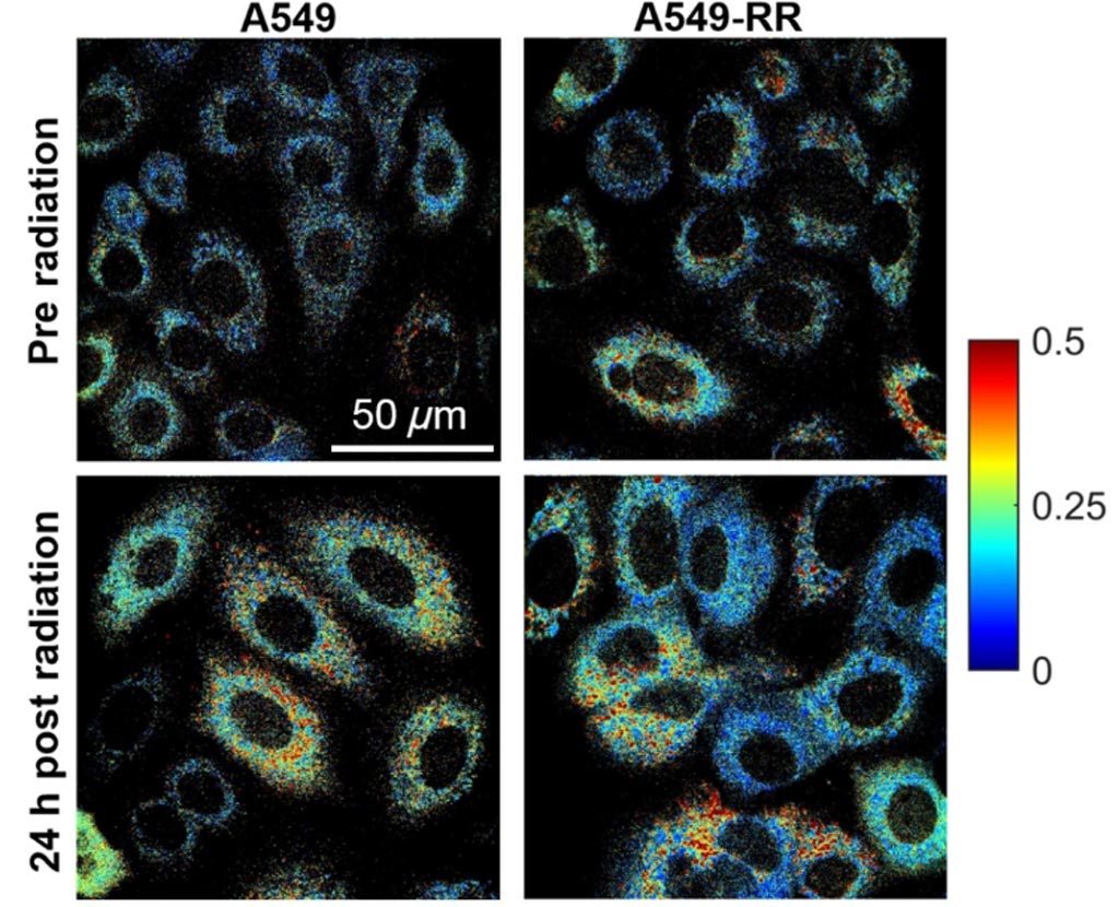
Image: Cell metabolism changes reflected in autofluorescence imaging (Photo courtesy of University of Arkansas).
By measuring the relative contributions of NADH and FAD to these processes, the researchers hoped to evaluate biochemical details related to cell metabolism. They therefore exposed the A549 cells to both radiation and the chemotherapy drug YC-1, a potent HIF-1α inhibitor. With the aid of fluorescence microscopy, they determined changes in the optical redox ratio of FAD/(NADH+FAD) over a period of 24 hours following treatment with YC-1, radiation, and both radiation and YC-1. They also evaluated changes in mitochondrial organization, glucose uptake, reactive oxygen species (ROS), and reduced glutathione.
They found significant differences in the optical redox ratio of radiation-resistant and sensitive A549 cells in response to radiation or YC-1 treatment alone; however, the combined treatment eliminated these differences. According to the researchers, the results show that optical redox ratio can reveal radiosensitization of previously radiation-resistant A549 cancer cells, and also provide a method for evaluating treatment response in patient-derived tumor biopsies. The study was published on June 11, 2018, in Scientific Reports.
“The use of autofluorescence imaging of cell metabolism can identify treatment-resistant cancer cells. More importantly, we think that this technique provides a sound method to evaluate tumor response to treatment and match tumors to the right therapy,” said senior author biomedical engineer Narasimhan Rajaram, PhD, of the University of Arkansas. “Non-destructive, label-free imaging approach is a valuable technique for characterizing metabolic reprogramming, and has potential clinical application to identify treatment efficacy in tumor-derived organoids.”
Hypoxic tumors tend to respond poorly to radiation because DNA damage relies on the presence of oxygen. As a regulator of oxygen homeostasis, HIF-1α plays a key role in downregulating mitochondrial oxygen consumption and enhancing transcription of important glycolytic genes; targeting HIF-1α has therefor shown promise in sensitizing radiation-resistant cancer cells to radiotherapy. Other factors in the tumor microenvironment, such as poor oxygen perfusion, can also contribute to elevated levels of HIF-1α, thereby compounding the cellular response to radiation.
Related Links:
University of Arkansas
University of Arkansas for Medical Sciences


