Fluoroscopy Bundle Curbs Radiotherapy Exposure
By MedImaging International staff writers
Posted on 05 Oct 2017
A system comprised of propriety software algorithm and hardware components helps address over exposure to radiation in hospital operating rooms (OR), particularly in the case of minimally invasive spine surgery.Posted on 05 Oct 2017
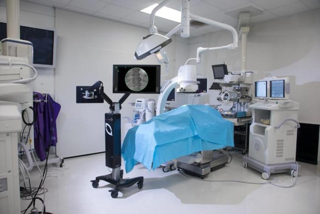
The NuVasive (San Diego, CA, USA) LessRay software system uses proprietary image enhancement technology in order to improve low-dose, low-radiation fluoroscopy (or x-ray) images, so that they have similar diagnostic capabilities as those of conventional full-dose fluoroscopy images, thus reducing radiation emission and exposure. Other features that reduce radiation exposure time include C-arm tracking, which simplifies localization and target anatomy quickly, accurately, and without the unnecessary scouting images. Economic benefits include no disruption to current surgical workflow and no additional surgery time needed.

Image: A novel system reduces fluoroscopy times during spine surgery (Photo courtesy of NuVasive).
An angle finder minimizes the number of OR steps required in order to obtain crisp endplate shots, with fewer fluoroscopic images than traditional C-arm scouting, and Alternate view, a feature that improves visualization by making metal instruments invisible or semi-transparent by fading out their obstruction. And image-stitching software minimizes surgical workflow interruption by rapidly stitching fluoroscopic images of any spine segment. A recent study demonstrated that radiation exposure of OR healthcare staff was 62-84% lower with LessRay, compared to conventional fluoroscopy.
“With LessRay's ability to dramatically reduce radiation exposure, as well as its enhanced imaging capability, we are now executing on our imaging and navigation strategy to improve spine surgery productivity, and ultimately predictability,” said Gregory Lucier, chairman and CEO of NuVasive. “Given the healthy pipeline of interest from customers already, the strong demand demonstrates a critical need for a solution like LessRay to address a major safety issue related to radiation facing surgeons and hospital administrators.”
“LessRay's ability to reduce radiation usage is so compelling because it benefits not only the surgeon but also the patient and everyone in the operating room,” said neurosurgeon Stephen Ryu, MD, of Palo Alto Medical Foundation (CA, USA). “Additional features that augment the capability of conventional fluoroscopy provide really useful tools for even the simplest surgeries. LessRay's capability to assist accurate localization of spine levels alone is worth it.”
Studies show spine and orthopedic surgeons can receive their lifetime occupational radiation limit within the first 10 years of their career. As a result, subsequent cancer rates and contraction of cataracts associated with radiation exposure are nearly double that of other surgery practices. Surgeons and support staff can use lead aprons and gloves, thyroid shields and protective glasses to try to minimize this, but this can have an impact on image quality, making it grainy and harder to interpret.
Related Links:
NuVasive


