PET Tracer Detects Thrombi in Blood Vessels and Brain
By MedImaging International staff writers
Posted on 17 Jul 2017
The results of a new pre-clinical study show that a fluorine-18 (18F) labeled ligand can be used to target GPIIb/IIIa receptors, and detect tiny blood clots during a diagnostic imaging scan.Posted on 17 Jul 2017
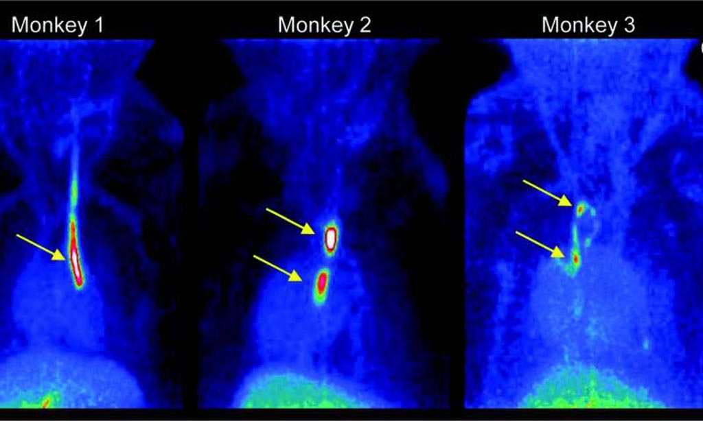
The small molecule tracer 18F-GP1 shows a high affinity for GPIIb/IIIa receptors, and accumulates at sites where blood clots are formed. The binding affinity of the novel Positron Emission Tomography (PET) tracer was not affected by heparin, aspirin and other anticoagulants, and was cleared rapidly from the blood stream.

Image: Strong signals are detected at the sites where inserted catheters had roughened surfaces. Almost no other background signal is visible. Only accumulation in the gallbladder becomes visible at the bottom of the image (Photo courtesy of Piramal Imaging).
The results were published in the July 2017 issue of The Journal of Nuclear Medicine’s (JNM) by the researchers from Piramal Imaging (Berlin, Germany). The researchers used Cynomolgus monkey models, and showed how the new technique could detect small arterial and venous blood clots, as well as emboli and endothelial damage in the brain.
The researchers are now working on a first-in-human study of 18F-GP1 and have presented the preliminary results, and an interim analysis that confirmed the results from the earlier preclinical data, at the annual Society of Nuclear Medicine and Molecular Imaging (SNMMI) meeting in June 2017.
Piramal Imaging researcher, Andrew W. Stephens, MD, PhD, said, “Currently available diagnostic techniques of thrombus [blood clot] imaging rely on different modalities depending on the vascular territory. A single imaging modality that could visualize thrombi from various sources in different anatomic regions would be very valuable. Although the current studies are preliminary, 18F-GP1 may provide not only more accurate anatomic localization, but also information of the risk of the clot growth or embolization. This may lead to changes in clinical intervention to the individual patient.”
Related Links:
Piramal Imaging


