Combined Imaging Technique Used in Breast Surgery
By MedImaging International staff writers
Posted on 13 Jun 2017
Researchers in the UK have shown that a combined optical and molecular imaging technique can be used to assess tumor margins during breast-conserving surgery.Posted on 13 Jun 2017
The researchers used Cerenkov Luminescence Imaging (CLI) together with a Positron Emission Tomography (PET) radiotracer F-18-fluorodeoxyglucose (F-18-FDG) for this first-in-human trial of the technique.

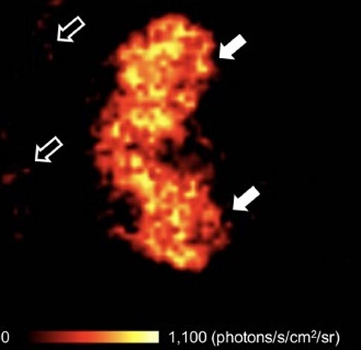
Image: By combining optical and molecular imaging, British researchers have developed a potential solution to help surgeons get the tumor margins right the first time during breast surgery (Photo courtesy of King’s College).
The study was carried out by researchers from King's College (London, UK) and published in the June 2017 issue of The Journal of Nuclear Medicine. Clinicians injected F-18-FDG tracer 45-60 minutes before surgery and then intraoperatively imaged tumor specimens in an investigational CLI imaging system immediately following tumor excision, for 22 patients suffering from invasive-breast cancer.
The results showed that F-18-FDG CLI was a useful low-risk tool for the intraoperative assessment of tumor margins during breast-conserving surgery.
Professor at King's College, Arnie D. Purushotham, MD, said, "Currently, approximately 1 in 5 women who undergo breast-conserving surgery, also known as lumpectomy, require repeat surgery due to inadequate excision of the tumor during the initial surgical procedure. By accurately assessing tumor resection margins intraoperatively with CLI, surgeons may be able to completely clear the cancer with a single operation, thereby reducing the number of breast cancer patients requiring a second, or even third, surgical procedure. The feasibility of intraoperative CLI as shown in this study, in combination with the wide applicability of F-18-FDG across a range of solid cancers, provides a stepping stone for clinical evaluation of this technology in other solid cancer types that also experience incomplete tumor resection due to close or involved margins."
Related Links:
King's College


