MR-Linac System for Difficult Cases Unveiled
By MedImaging International staff writers
Posted on 16 May 2017
An innovative new MR/RT system that could change cancer care especially for cases that are difficult to treat has been unveiled.Posted on 16 May 2017
The manufacturer expects the system to go on sale in Europe and globally, later in 2017, when it is expected to receive the CE Mark (Conformité Européenne).

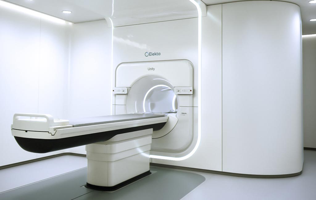
Image: The Unity MR-linac system was unveiled at the ESTRO 36 meeting in Vienna, Austria (Photo courtesy of Elekta).
The Elekta Unity was developed by Elekta and is the only Magnetic Resonance Radiation Therapy (MR/RT) system that combines a premium diagnostic quality (1.5T) MRI scanner with an advanced linear accelerator (linac). The system is designed to deliver a precise radiation dose, and at the same time capture high-quality MR images. This will enable clinicians to visualize tumors, and adapt treatment. Unity features a compact, 70 cm wide-bore MRI, a table with a low load height for patient comfort and a soft tabletop, and non-glare room lighting.
Unity is the fruit of a global partnership consortium set up by Elekta with Royal Philips as MR technology partner. The consortium has also developed new clinical and workflow protocols for the system.
Conceptual architect and inventor of the system, Professor Jan Lagendijk, PhD, Head Radiation Oncology, University Medical Center Utrecht, said, “We started the development of an integrated MR-linac system 18 years ago; the presentation of the clinical system is a huge milestone. This system will enable tumor dose escalation and extreme normal tissue sparing, it will also enable shorter and more effective treatment regimens, while its ability to perform functional imaging has the potential to perform better dose painting and tumor response assessment.”


