Microscopic Sensor Helps Tailor Radiology Treatments
By Daniel Beris
Posted on 23 Nov 2016
A new sensor measures radiation doses at the individual cell level, enabling clinicians to obtain a complete picture of how much damage each cell has incurred following treatment.Posted on 23 Nov 2016
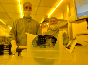
Developed by researchers at SINTEF (Trodheim, Norway), the University of Wollongong (UOW; Australia), and other institutions, the new sensor is as small as a cancer cell, a feat achieved by using a technology called semiconductor processing. The measuring instrument thus contains a sheet of microsensors placed side-by side, all mounted on a silicon base. The sensors are also encapsulated in a plastic material that mimics human tissue, so that the radiation dose measured by the sensors is almost identical to that absorbed by real cancer cells.

Image: Researchers Marco Povoli and Angela Kok with the microscopic radiation sensor (Photo courtesy of SINTEF).
The dispersal of mixed radiation fields across a given area enables the sensors to provide an image of the location within the cell that absorbs the highest levels of radiation. The sensor was recently tested at the European Synchrotron Radiation Facility (ESRF; Grenoble, France), and was found to be capable of measuring true radiation doses absorbed by tissue, and with a better spatial resolution than existing equipment. The researchers are now working to provide more precise quantification of the radiation doses absorbed by cancer tissue, and reduce the damage incurred by healthy tissue.
“The most important component of the new sensor is the element silicon, which is a semiconductor with radiation detection properties. When radiation counteracts with silicon the energy is converted into a measurable electrical signal. The magnitude of the signal indicates the intensity of the radiation,” said SINTEF researcher physicist Angela Kok, PhD. “This technology means that doctors can monitor and control radiation doses to make sure that only cancer cells are destroyed, with only minimal damage to surrounding healthy tissue.”
“It appears that proton therapy produces better outcomes for some types of cancer than traditional radiotherapies. There currently exist no sensors capable of measuring radiation of this kind, but we realized that our technology could be adapted to develop sensors with the right specifications,” added SINTEF research scientist Marco Povoli, PhD. “The fabrication process required more development to optimize the reliability of the results, but we overcame this challenge within a few months.”
Related Links:
SINTEF
University of Wollongong
European Synchrotron Radiation Facility


 Guided Devices.jpg)


