Dedicated CT Scanner Advances Radiation Therapy Simulation
By MedImaging International staff writers
Posted on 12 Oct 2016
A new computerized tomography (CT) scanner provides personalized images for radiation therapy (RT) treatment planning.Posted on 12 Oct 2016
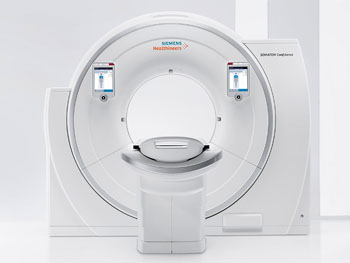
The Siemens Healthineers (Erlangen, Germany) Somatom Confidence RT Pro CT scanner provides all the functionalities of a CT simulator, which produces images that are used by physicians to define the target and organs at risk for treatment prescription. The device comes with the new DirectDensity algorithm, which provides electron density images that allow physicians to contour treatments on personalized images acquired with individualized kV settings. At the same time physicists get the physical property readings they need to perform dose calculations, all in a simplified workflow.

Image: The Somatom Confidence RT Pro CT scanner (Photo courtesy of Siemens Healthineers).
Another feature is iMAR, an algorithm that removes metallic artifacts due to implants and orthopedic fixations. Currently, physicists manually correct for artifacts prior to dose calculation, while physicians often guess while contouring. iMAR can significantly reduce these operational inefficiencies, improving quality of care. Another feature is Dual Energy capabilities; the Somatom Confidence RT automatically acquires two CT scans at different kV voltages, which can then be manipulated to improve visualization.
The Somatom Confidence RT Pro is complemented by the new syngo.via RT Image Suite, a multimodality simulation and advanced contouring software solution that streamlines the simulation process. In addition, it helps determine the right treatment strategy for moving tumors--such as those found in the liver and lungs--by visualizing quantitative three dimensional (3D) tumor trajectories. It also facilitates the adoption of new treatment approaches such as mid-ventilation, which can increase the number of patients potentially eligible for stereotactic body radiation therapy (SBRT).
“This new offering complements the dedicated Siemens Healthineers MR, CT, PET/CT, and software portfolio for radiation therapy, and demonstrates how it can help healthcare facilities improve their outcomes while also lowering costs,” said Gabriel Haras, MD, head of radiation oncology at Siemens Healthineers.
During treatment preparation, radiation oncologists need a quality CT image for contouring tumors and sparing healthy-organ tissue, while physicists need a CT image that shows the patient's electron density for treatment planning purposes. Currently, CT images for RT were optimized primarily for electron density representations, which dictated fixed tube voltage and kV settings, limiting CT scanners from realizing their full potential and capabilities.
Related Links:
Siemens Healthineers


