PET/CT Better Choice Than Bone Marrow Biopsy for Diagnosis, Prognosis of Lymphoma Patients
By MedImaging International staff writers
Posted on 13 Aug 2013
A more accurate technique for determining bone marrow involvement in patients with diffuse large B-cell lymphoma (DLBCL) has been identified by French researchers. Posted on 13 Aug 2013
18F-fluorodeoxyglucose (FDG) positron emission tomography/computed tomography (PET/CT) imaging when compared to bone marrow biopsy, was found to be more sensitive, demonstrated a higher negative predictive value, and was more accurate for diagnosing these patients— changing treatment for 42% of patients with bone marrow involvement.

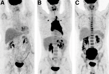
Image: Diffuse bone marrow uptake pattern in 18F-FDG PET/CT. (A and B) Uptake lower than (A) or similar to (B) that in liver was considered negative for BMI. (C) Uptake higher than that in liver was always linked to anemia or inflammatory processes and also considered negative for BMI (Photo courtesy of the Society of Nuclear Medicine and Molecular Imaging).
DLBCL is the most frequent subtype of high-grade non-Hodgkin lymphoma, accounting for nearly 30% of all newly diagnosed cases in the United States. In recent decades, there has been a 150% increase in incidence of DLBCL. “In our study, we showed that in diffuse large B-cell lymphoma, 18F-FDG PET/CT has better diagnostic performance than bone marrow biopsy to detect bone marrow involvement and provides a better prognostic stratification. While bone marrow biopsy is considered the gold standard to evaluate bone marrow involvement by high-grade lymphomas, 18F-FDG PET/CT is in fact the best method to evaluate extension of the disease, as well as avoid invasive procedures,” said Louis Berthet, MD, from the Centre Georges-Francois Leclerc (Dijon, France), and lead author of the study, which was published in the August 2013 issue of the Journal of Nuclear Medicine.
The retrospective study included 133 patients diagnosed with DLBCL. All patients received both a whole-body 18F-FDG PET/CT scan, as well as a bone marrow biopsy to determine bone marrow involvement. A final diagnosis of bone marrow involvement was made if the biopsy was positive, or if the positive PET/CT scan was confirmed by a guided biopsy, by targeted magnetic resonance imaging (MRI) or, after chemotherapy, by the concomitant disappearance of focal bone marrow uptake and uptake in other lymphoma lesions on 18F-FDG PET/CT reassessment. Progression-free survival and overall survival were then analyzed.
Thirty-three patients were considered to have bone marrow involvement. Of these, eight were positive according to the biopsy and 32 were positive according to the PET/CT scan. 18FDG PET/CT was more sensitive (94% vs. 24%), showed a higher negative predictive value (98% vs. 80%) and was more accurate (98% vs. 81%) than bone marrow biopsy. Among the 26 patients with positive 18F-FDG PET/CT results and negative biopsy results, 11 were restaged to stage IV by PET/CT, which changed their treatment plans.18F-FDG PET/CT was also determined to be an independent predictor of progression-free survival.
“Our findings add to the literature to prove the significance of 18F-FDG PET/CT in cancer evaluation and to democratize this imaging method,” concluded Dr. Berthet. “Molecular imaging is the best method to adapt targeted therapies to each patient. The emergence of PET/MRI and novel radiotracers predicts an exciting new future for our field.”
Related Links:
Centre Georges-Francois Leclerc


 Guided Devices.jpg)


