Stereotactic Radiosurgery Effective Within Standard Time Slots
By MedImaging International staff writers
Posted on 16 May 2012
A highly precise and noninvasive way of excising tumors using carefully shaped high-energy X-ray beams can be performed quickly and accurately in the treatment of tumors of the brain, spine, thorax, and gastrointestinal (GI) tract. Posted on 16 May 2012
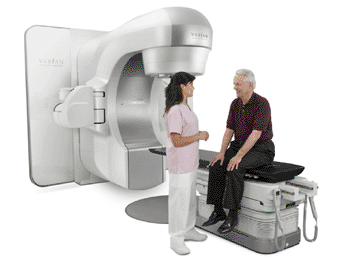
The Varian TrueBeam STx system was designed for efficient, precise radiosurgery, delivering treatments two to eight times faster than other radiosurgery systems when operating in high intensity mode. Intelligent automation further speeds treatments with an up to five-fold reduction in the number of steps needed for imaging, positioning, and treating patients, compared with earlier generations of technology. Additionally, a high-definition multileaf collimator shapes the radiation dose so that it closely matches the shape of the targeted tumor, in order to precisely carry out delicate procedures near critical structures such as the optic chiasm, the brain stem, or the spinal cord.

Image: The Varian TrueBeam STx system (Photo courtesy of Varian Medical Systems).
On a TrueBeam system, sophisticated RapidArc radiosurgery procedures for spinal and brain lesions can be carried out within a standard 15-minute radiotherapy time slots, according to a recent study carried out by researchers from the University of Alabama (UAB; Birmingham, USA). These are treatments that can take 45 minutes to an hour or longer using standard cobalt-based or robotic technology systems. The Varian TrueBeam STx system is a product of Varian Medical Systems (Palo Alto, CA, USA).
“Linear accelerator radiosurgery has come a long way, matching and even surpassing what was possible with earlier generations of radiosurgery technology,” commented Antonio De Salles, MD, PhD, a professor of neurosurgery at the University of California, Los Angeles (UCLA, USA). “It has been exciting to see these capabilities evolve along with the technology, and become applicable for treating lesions not only inside the cranium, but in the rest of the body as well.”
Related Links:
Varian Medical Systems
University of Alabama
University of California, Los Angeles


