Endoscopic Molecular-Guided Surgery Eradicates Cancer
By MedImaging International staff writers
Posted on 09 Apr 2012
New luminescent endoscope technology shows promise helping surgeons remove cancerous tumors more completely.Posted on 09 Apr 2012
Developed by researchers at Stanford University (CA, USA), the procedure known as Cerenkov Luminescence Endoscopy (CLE), holds advantages over both traditional endoscopic and imaging techniques such as magnetic resonance imaging (MRI), by providing information about the functioning of the tissue itself. CLE is a development of Cerenkov Luminescence Imaging (CLI), which can produce images of organs, and guide surgery to remove remaining cancer cells that otherwise would have been invisible to surgeons.

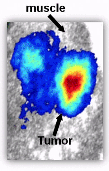
Image: CLI technology demonstrates the presence of a tumor (Photo courtesy of Zhen Cheng, PhD).
CLE and CLI rely on a physical phenomenon known as Cerenkov radiation, an electromagnetic radiation emitted when a charged particle (such as an electron) passes through a dielectric medium at a speed greater than the phase velocity of light in that medium; it is responsible for the soft blue glow in the cooling water in the core of nuclear power reactors. This luminescence is especially exciting, because the light used to reveal diseased tissue is in the visible spectrum, and can be detected with simple optical sensors. The technology was presented at the 243rd National Meeting & Exposition of the American Chemical Society (ACS), held during March 2012 in San Diego (CA, USA).
“The weak blue light -- unlike the X-rays in other medical scans -- barely penetrates through deep tissues. This limits the usefulness of the technology in humans, where many tumors develop in areas deep inside the body,” said lead researcher Zhen Cheng, PhD. “Our marriage of Cerenkov luminescence with the endoscope may be the perfect solution. With endoscopy, we can get close enough to the diseased tissue to take advantage of this technology.”
CLI has also been found to dramatically improve the resolution of positron emission tomography (PET) scans, enabling PET scanners to detect smaller objects than previously possible.
Related Links:
Stanford University


 Guided Devices.jpg)


