Novel PET Technique Visualizes Spinal Cord Injuries to Predict Recovery
Posted on 24 Feb 2025
Each year, around 18,000 individuals in the United States experience spinal cord injuries, leading to severe mobility loss that often results in a lifelong battle to regain independence and improve quality of life. In the initial months following an injury, there are rapid degenerative changes, including demyelination at the site and surrounding areas of the lesion, which can lead to less favorable recovery outcomes. The prognosis for recovery following spinal cord injuries is often unpredictable, and current imaging techniques do not always offer clear insights into which patients may recover. Now, a novel PET imaging technique for visualizing spinal cord injuries has now shown promise in providing critical information on which patients may regain mobility, according to recent research published in The Journal of Nuclear Medicine.
A team of researchers from Spaulding Rehabilitation Hospital (Charlestown, MA, USA) and Massachusetts General Hospital (Boston, MA, USA) conducted a study to determine whether PET imaging could identify spared nerve connections within the injured spinal cord that might indicate a better likelihood of recovery. To achieve this, a new PET tracer, 18F-3F4AP, was developed to target and visualize demyelinated axons, or damaged nerve fibers, in spinal cord injuries. In a preclinical study, rats with incomplete spinal cord contusion injuries were imaged using 18F-3F4AP PET at various time points, up to a month after the injury. The PET results were then compared with autoradiography and immunohistochemistry of postmortem spinal cord tissue. Additionally, a proof-of-concept study was conducted on two human patients with spinal cord injuries of varying severities using the 18F-3F4AP PET technique.
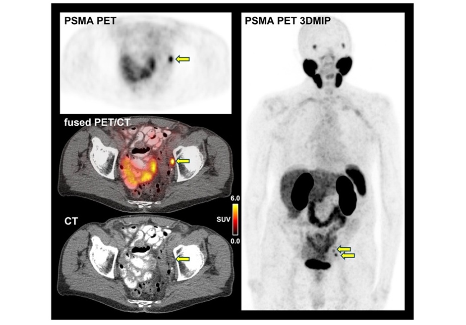
![Image: [18F]3F4AP in a human subject after mild incomplete spinal cord injury (Photo courtesy of The Journal of Nuclear Medicine, DOI:10.2967/jnumed.124.268242)](https://globetechcdn.com/mobile_medicalimaging/images/stories/articles/article_images/2025-02-24/Brugarolas_F8.large.jpg "Image: [18F]3F4AP in a human subject after mild incomplete spinal cord injury (Photo courtesy of The Journal of Nuclear Medicine, DOI:10.2967/jnumed.124.268242)")
The 18F-3F4AP PET scans of the rats revealed a more than two-fold increase in tracer binding at the injury site seven days post-injury compared to baseline. Autoradiography, histology, and immunohistochemistry further validated that 18F-3F4AP targeted demyelinated axons. In human studies, 18F-3F4AP PET successfully differentiated between severe spinal cord injuries and injuries with significant recovery, revealing axonal loss in the former and demyelination in the latter. Furthermore, dynamic PET images displayed alterations in tracer delivery, indicating differences in spinal cord blood flow between the two injury types.
“This study underscores the incredible potential of molecular imaging to investigate complex neurological challenges, such as spinal cord injuries, multiple sclerosis, traumatic brain injury, and even some dementias,” said Pedro Brugarolas, PhD, assistant professor at Massachusetts General Hospital. “Such a tool could revolutionize how we diagnose, monitor, and understand neurological diseases, paving the way for advancements in molecular imaging and nuclear medicine.”