New Technique Combines X-Ray Imaging and Radar for Safer Cancer Diagnosis
Posted on 08 Aug 2025
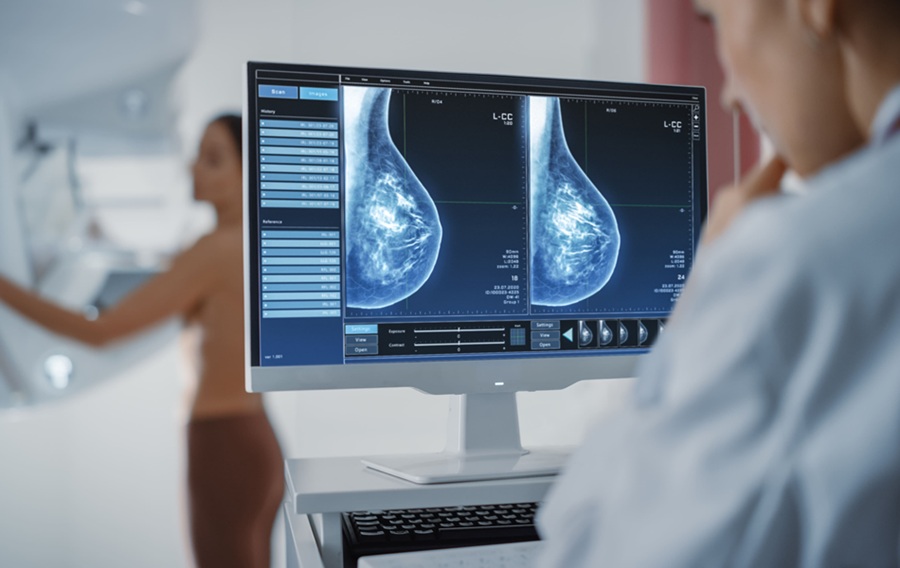
X-ray imaging methods, such as mammography and computed tomography (CT) scans, are essential for diagnosing and monitoring breast and lung cancer. While CT provides valuable three-dimensional insights, it comes with significant drawbacks due to its high radiation dose—around three times the amount of natural annual exposure—posing health risks to patients. This is especially concerning in cancer care, where repeated imaging is often required. As a result, there is a need for methods that deliver accurate and detailed diagnostic information with reduced radiation exposure. Now, a new solution aims to address this issue by combining two imaging modalities to improve diagnostic accuracy while significantly reducing the burden on patients.
As part of the MultiMed project, researchers at Fraunhofer-Gesellschaft (Freiburg, Germany) are creating a multimodal lab system that integrates radar with X-ray CT imaging. Radar, though more commonly used in aviation and automotive industries, can also produce three-dimensional medical images without exposing patients to radiation. It detects changes in tissue by measuring electrical permittivity and conductivity. The team has developed methods to co-register radar and X-ray data—placing both datasets in spatial relation to one another.
")
Additionally, the researchers are enhancing radar reconstruction algorithms to improve image clarity and integrating radar data into the X-ray CT reconstruction process. This multimodal CT algorithm boosts the quality and detail of CT images, reduces artifacts, and lowers radiation exposure. The researchers have created imaging phantoms—realistic artificial models of tissue structures—to test the method. These phantoms allow the system to simulate real diagnostic conditions and confirm that both radar and X-ray signals deliver meaningful results. Once completed, this system could offer a new, patient-friendly way to detect cancer-related tissue changes early and accurately while minimizing radiation exposure.
"The new approach has the potential to detect tissue changes early on and with great accuracy — and with much less of a burden on patients than before," said Victoria Heusinger-Hess, Project Manager, Fraunhofer Institute for High-Speed Dynamics.
Related Links:
Fraunhofer-Gesellschaft