New Detection Method Spares Unnecessary Prostate Cancer Treatments
By MedImaging International staff writers
Posted on 18 Apr 2016
A novel three-dimensional (3D) cell model that mimics the morphogenesis of acini, the glandular units that constitute the prostate, could help study how prostate cancer (PC) progresses.Posted on 18 Apr 2016
Developed by researchers at St. George's University of London, the 3D cellular model monitors the morphogenesis of the glandular structures using microscopy and imaging techniques, which can help identify the intraluminal hyperproliferation of the acini. By identifying the factors that alter the epithelial organization of acini that lead to the intraluminal proliferation, the researchers hope to develop a strategy that can help determine the causes of PC progression.
.")
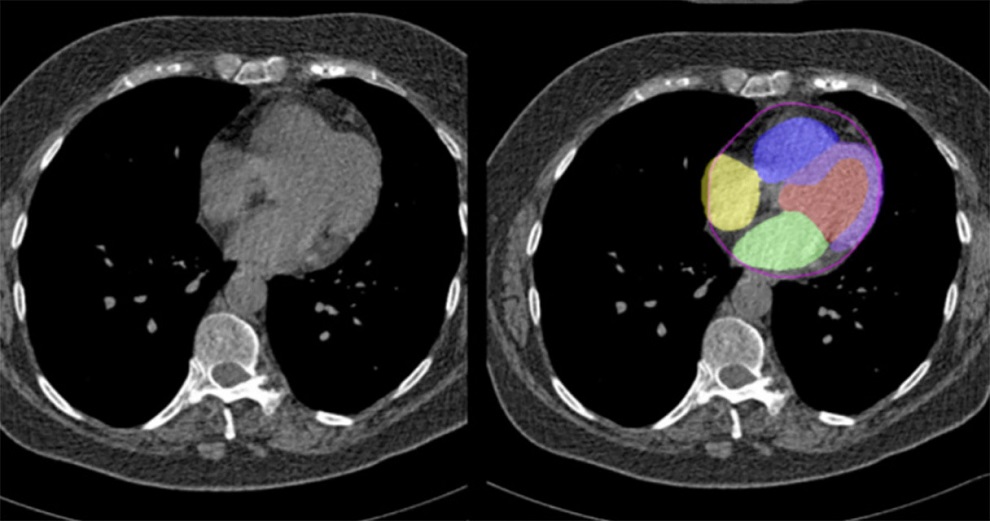
Scanning for prostate cancer for 3D modeling (Photo courtesy of St. George\'s University of London).
Using the 3D acini model, the researchers are developing methods that will provide objective measures for patient stratification and disease grading, which could thus complement current clinical classification of patients diagnosed with PC. By doing so, they hope to be able to consistently distinguish aggressive PC from indolent disease, a distinction currently difficult to identify, which often leads to inappropriate treatment and high morbidity and mortality.
“Based on these cell culture models, we believe a test could be developed to assess how invasive a prostate cell could become when taken from a patient,” said principal investigator Ferran Valderrama, PhD, of the SGUL PC cell biology laboratory. “We believe that this information would help to determine the appropriate treatment for the patient, reducing the burden derived from unnecessary over treatment, and overall having a positive outcome in patient survival.”
“The currently available prognostic tests for prostate cancer cannot conclusively tell us whether a tumor will rapidly progress and spread to other tissues, or instead will remain confined to the limits of the prostate,” added Dr. Ferran Valderrama. “Improving current procedures for predicting the outcome of a prostate that presents with ‘suspicious’ characteristics of prostate cancer are necessary. If we were able to determine that outcome in a consistent and conclusive manner, it would be easier to define the best approach for treatment for each individual case.”
Related Links:
St George's University of London

Gold Member
Solid State Kv/Dose Multi-Sensor
AGMS-DM+

Color Doppler Ultrasound System
DRE Crystal 4PX

New
X-Ray QA Meter
Piranha CT

New
Mobile Digital C-arm X-Ray System
HHMC-200D