New 3D Imaging System Addresses MRI, CT and Ultrasound Limitations
Posted on 20 Jan 2026
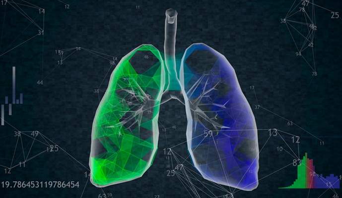
Medical imaging is central to diagnosing and managing injuries, cancer, infections, and chronic diseases, yet existing tools each come with trade-offs. Ultrasound, X-ray, CT, and MRI can be costly, time-consuming, limited in resolution or depth, or rely on ionizing radiation or strong magnets. These constraints make it difficult to rapidly capture detailed images across large areas of the body. To address these challenges, researchers have demonstrated a non-invasive imaging approach that can quickly generate three-dimensional views of both tissue structure and blood vessels across wide regions of the human body, offering a potential new option for fast, comprehensive diagnostics.
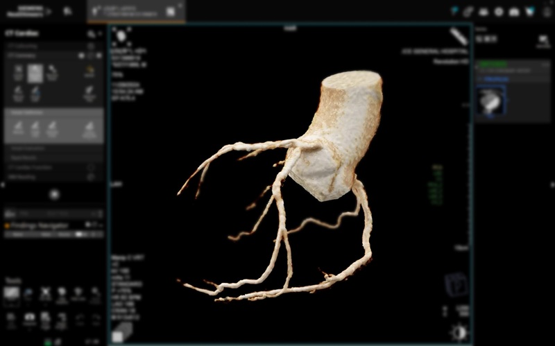
The technology, developed by researchers at the Keck School of Medicine of USC (Los Angeles, CA, USA), in collaboration with the California Institute of Technology (Caltech, Pasadena, CA, USA), overcomes key limitations of conventional imaging by combining complementary techniques into a single platform. The system integrates rotational ultrasound tomography with photoacoustic tomography. Ultrasound provides structural information about tissues, while photoacoustic imaging detects sound waves generated when laser light is absorbed by blood, allowing vessels to be visualized without contrast agents.
")
Unlike standard ultrasound, which typically produces 2D images, the new system uses an arc of detectors to reconstruct full 3D volumes. At the same time, laser pulses interact with hemoglobin in blood, generating ultrasonic signals that map vascular networks using the same detectors. This combined approach enables simultaneous imaging of soft tissue and blood vessels at meaningful depths, without exposing patients to radiation or requiring long scan times, and at a lower expected cost than MRI.
In a proof-of-concept study funded by the National Institutes of Health, the researchers tested the system in humans by imaging the brain, breast, hand, and foot. Brain imaging was performed in patients undergoing surgery for traumatic brain injury, where sections of the skull had been temporarily removed. The system captured detailed 3D images across regions up to 10 centimeters wide in about 10 seconds. The findings, published in Nature Biomedical Engineering, showed that the technique could reliably visualize both tissue architecture and blood vessels across multiple anatomical sites.
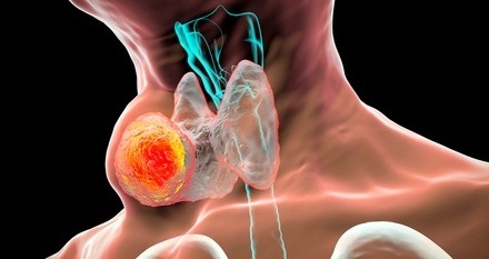
By demonstrating performance across different parts of the body, the study highlights the platform’s broad clinical potential. Applications could include brain imaging for stroke or traumatic injury, breast imaging for cancer care, and rapid vascular assessment in limbs affected by diabetes or venous disease. Further work is needed before routine clinical use, particularly to overcome signal distortion caused by the intact human skull. The team is now refining the system, including optimizing ultrasound frequencies and improving image consistency, as they move toward future clinical translation.
“You cannot understate the importance of medical imaging for clinical practice. Our team has identified key limitations of existing techniques and developed a novel approach to address them,” said Charles Liu, MD, PhD, co-senior author of the research. “When we think about the critical limitations of current medical imaging, including expense, field of view, spatial resolution and time to scan, this platform addresses many of them.”
Related Links:
Keck School of Medicine of USC
Caltech


 Guided Devices.jpg)