10-Minute Brain Scan Predicts Effectiveness of Spinal Cord Surgery
Posted on 04 Dec 2024
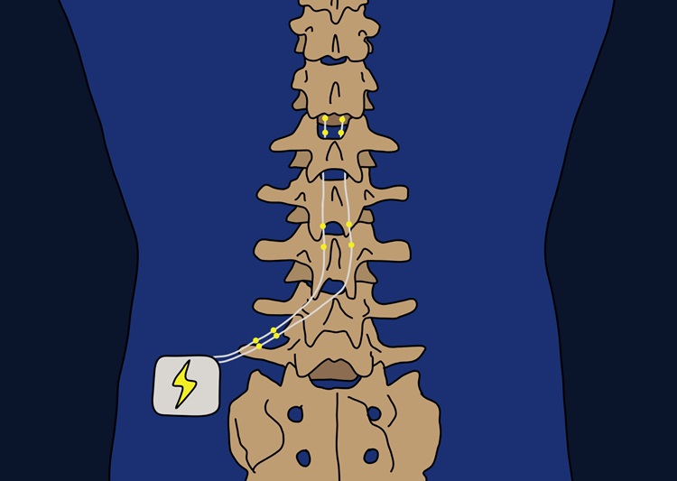
For patients suffering from chronic pain that cannot be alleviated through other treatments, spinal cord stimulation is often considered a last-resort solution. This procedure involves implanting leads into the spine and using electrical stimulation to interfere with pain signals sent to the brain. The leads are strategically placed so that patients experience the sensation of stimulation at the site of pain, as the spinal cord transmits sensations from various body parts. However, spinal cord stimulation doesn’t work for everyone, and the procedure’s effectiveness is typically assessed through a short trial lasting anywhere from a few days to two weeks before permanent implantation. While brief, this trial remains an invasive procedure with potential risks. As a result, clinicians have long sought non-invasive methods to predict which patients will benefit from this procedure. Now, a 10-minute brain scan has been shown to predict how well spinal cord stimulation will alleviate pain, offering doctors a valuable tool for discussing potential treatment outcomes with patients.
Functional magnetic resonance imaging (fMRI) has become a common technique for observing brain activity. It helps to identify which areas of the brain are activated by specific stimuli, providing insights into how different brain regions are connected. In an earlier study, researchers from Kobe University (Kobe, Japan) found that the effectiveness of pain relief from ketamine correlated with the strength of the connection between two areas of the default mode network before the drug was administered. This network, which plays a significant role in self-referential thoughts, has been linked to chronic pain. Another important factor is the connection between the default mode network and the salience network, which governs attention and responses to stimuli. The researchers decided to investigate whether the activity within and between these networks could be used to predict a patient’s response to spinal cord stimulation. The study involved 29 patients suffering from various forms of chronic pain that could not be easily treated.

In the study results published in British Journal of Anesthesia, the researchers found that patients who had better responses to spinal cord stimulation showed weaker connections between specific regions of the default mode network and the salience network. This discovery not only provides a potential biomarker for predicting treatment outcomes, but it also supports the hypothesis that disrupted connections between these networks may contribute to the development of chronic pain in the first place. While fMRI scans offer one method of assessment, the study also suggests that combining pain questionnaires and other clinical indicators can serve as a reliable alternative for predicting how well a patient will respond to spinal cord stimulation. The researchers argue that, despite the cost of MRI scans being debated, the burden on patients and healthcare providers could be significantly reduced if the effectiveness of spinal cord stimulation could be predicted with a single 10-minute resting state fMRI scan.
“We believe that more accurate evaluation will become possible with more cases and more research in the future,” said Kobe University anesthesiologist Kyohei Ueno who led the research team. “We are also currently conducting research on which brain regions are strongly affected by various patterns of spinal cord stimulation. At this point, we are just at the beginning of this research, but our main goal is to use functional brain imaging as a biomarker for spinal cord stimulation therapy to identify the optimal treatment for each patient in the future.”


