Accelerated MRI Sequence Helps Radiologists Assess Heart Disease without Breath-Holding
Posted on 14 Feb 2024
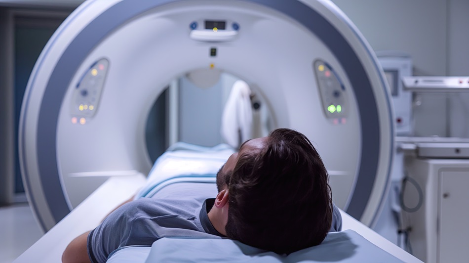
Cardiac magnetic resonance imaging (MRI) is essential for evaluating and diagnosing heart damage resulting from poor blood flow. Traditional cardiac MRI examinations are time-consuming and typically require patients to perform multiple breath holds to prevent respiratory artifacts. This can be challenging, especially for patients with ischemic heart disease, and may lead to compromised image quality and potential measurement inaccuracies. Now, an accelerated MRI sequence can help radiologists assess ischemic heart disease without requiring patients to hold their breath.
Scientists at Amiens University Hospital (Amiens, France) conducted a prospective study involving patients undergoing cardiac MRI for ischemic heart disease assessment between March and June 2023. The study included an innovative, free-breathing, short-axis imaging sequence that utilized deep learning for image reconstruction. Two radiologists evaluated the image quality of both the traditional and the new MRI methods. The study involved a total of 26 patients.

The results showed that the acquisition time was shorter with the deep learning-based method compared to the standard sequence. Furthermore, this accelerated approach demonstrated no significant difference in assessing the left ventricular ejection fraction, an important measure of heart function. The radiologists observed that the subjective image quality of the deep learning-based images was superior. However, they also noted an increased frequency of blurring artifacts in these images.
“The improved subjective image quality of the cine-deep learning sequence likely in part relates to the sequence’s respiratory synchronization, in comparison with the standard sequence’s reliance on adequate breath-holding, which may be challenging in dyspneic patients,” concluded David Monteuuis, MD, at Amiens University Hospital.
Related Links:
Amiens University Hospital


