World-First Benchmark for Measuring Brain Atrophy Created Using `Fake` MRIs Developed via AI
Posted on 08 Feb 2023
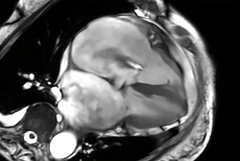
Alzheimer’s is the most common form of dementia and accounts for 60% to 80% of cases. One way to measure its progress is via magnetic resonance imaging (MRI) images that show cortical thinning. However, assessing the onset and progression of Alzheimer’s using brain MRI poses a challenge as changes in the thickness of the brain's cortex are extremely small, usually in the sub-millimeter range. Advanced machine learning techniques are generally used for brain research to examine changes in cortical thickness, although the absence of a clinically accurate ‘ground truth’ dataset meant that their sensitivity to the detection of small atrophy levels could not be evaluated. Until now, the only way to obtain a ground truth measure of cortical thickness was by studying the brain post-mortem. However, this again poses a challenge as the brain begins to shrink immediately after death, resulting in inaccurate readings.
Now, scientists from CSIRO (Canberra, Australia), in partnership with Queensland University of Technology (Brisbane, Australia), have used artificial intelligence (AI) to develop a world-first benchmark for measuring brain atrophy – or thinning - in neurodegenerative diseases, including Alzheimer’s disease. Cortical atrophy – thinning of the brain’s cortex – can begin up to 10 years before the appearance of clinical symptoms of Alzheimer’s disease. The new technique allows researchers to set the amount and location of brain degeneration they wish to compare against in order to achieve a clear picture of the best method for cortical thickness quantification. The technique can test the sensitivity of methods to a miniscule level and determine if a method can detect changes in thickness of just 0.01 millimeters.
")
The scientists believe they have strong evidence that DL+DiReCT – a deep learning-based method for measuring cortical thickness – is robust and sensitive to subtle changes in atrophy. The technique can be applied to research in any brain disease involving neurodegeneration and marks a significant step forward in better understanding dementia and other debilitating brain diseases. The technique could also be used to predict the level of cortical degeneration expected in a person over time. The technology was developed on the back of the commonly used and relatively inexpensive MRI images. The researchers have made the synthetic dataset images publicly available for clinicians and scientists who can use the synthetic images to perform their own assessments of cortical thickness quantification methods.
“Using the power of machine learning, we were able to produce a set of artificial MRI images of brains with predefined signs of neurodegeneration in the cortex region, the outer layer of the brain most affected by Alzheimer’s,” said Filip Rusak, research scientist from CSIRO’s Australian e-Health Research Centre. “Before these findings, there was no way to conclusively determine the sensitivity of the various methods used to measure cortical thickness in Alzheimer’s patients.”
Related Links:
CSIRO
Queensland University of Technology