MRI-Based Technology Assesses Muscle Composition
By MedImaging International staff writers
Posted on 21 Dec 2021
Novel software can analyze a patient with suspected sarcopenia using a rapid neck-to-knee magnetic resonance imaging (MRI) scan.Posted on 21 Dec 2021
The AMRA medical (Linköping, Sweden) Muscle Assessment Score (MAsS) Scan is designed to measure volumetric changes in muscle mass and the diffusion of fat infiltration into the muscle. The scanning protocol is based on symmetrical chemical shift imaging, also known as two-point Dixon imaging. Acquired images consist of fat and water image pairs, as reconstructed by the software. The images are automatically calibrated and corrected for variations caused by inhomogeneity in the magnetic field and coil sensitivity.

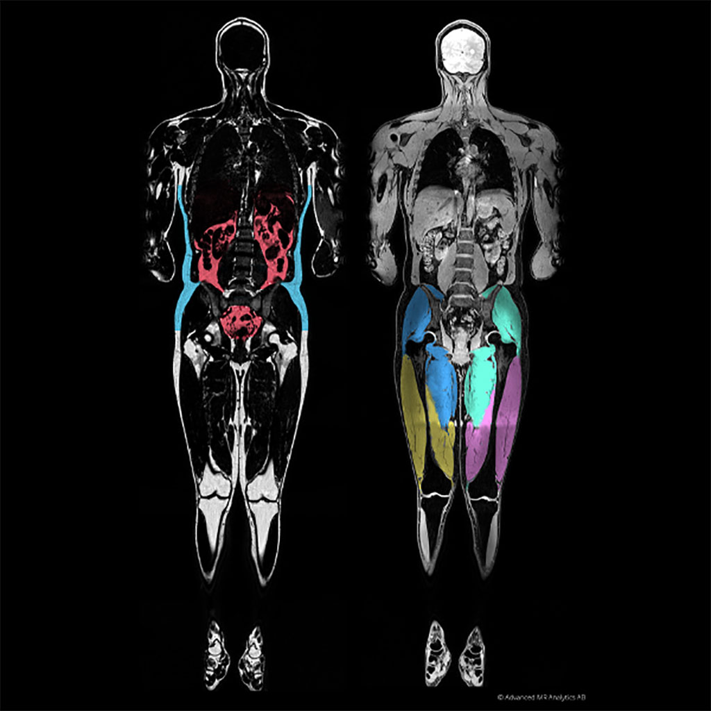
Image: A graphical MAsS score of visceral fat (L) and muscle (R) (Photo courtesy of AMRA medical)
The resulting report contains the MAsS scan, anatomic and color-coded images, and precise body composition measurements with contextual insights based on AMRA's reference database. The platform-agnostic software works across all major 1.5 and 3T MR scanners, such that output across scanners is standardized and calibrated using the fat signal as an internal reference. The value of each pixel shows the percentage of fat in it; partial-volume effects do not affect quantification, and thin layers of fat (or even diffuse infiltration) contribute to fat quantification.
In addition, AMRA automates the classification and quantification of fat and muscle groups, with segmentation based on registration between the image data volume and manually segmented prototype volumes. Body fat is divided into subcutaneous, visceral, and ectopic compartments; muscle groups are automatically classified, and the volume of each individual muscle group is obtained. Additionally, the amount of fat in any user-defined region, (e.g. a muscle or an internal organ) can be calculated also for diffuse fat infiltration.
“The AMRA MAsS Scan will greatly benefit patients by allowing clinicians to assess sarcopenia and improve patient outcomes an objectively and accurately assess muscle quality and take action,” said Eric Converse, CEO of AMRA Medical. “The beauty of the report is that it is easy-to-understand, it creates a common language among clinicians with the muscle assessment score, and adds only minutes to an already prescribed MRI.”
Sarcopenia (from the Greek, meaning "poverty of flesh") is the degenerative loss of skeletal muscle mass. Sarcopenia is characterized first by muscle atrophy, along with a reduction in muscle tissue "quality," caused by such factors as replacement of muscle fibers with fat, an increase in fibrosis, changes in muscle metabolism, oxidative stress, and degeneration of the neuromuscular junction. Combined, these changes lead to progressive loss of muscle function and frailty. Sarcopenia can be thought of as a muscular analog of osteoporosis, also caused by inactivity and counteracted by exercise. The combination of osteoporosis and sarcopenia results in the significant frailty often seen in the elderly population.
Related Links:
AMRA medical


 Guided Devices.jpg)


