MRI Helps Create Personalized Brain Temperature Maps
By MedImaging International staff writers
Posted on 20 May 2021
A new study shows how data from magnetic resonance imaging (MRI) of brain tissue and vessel structure can facilitate personalized brain temperature predictions. Posted on 20 May 2021
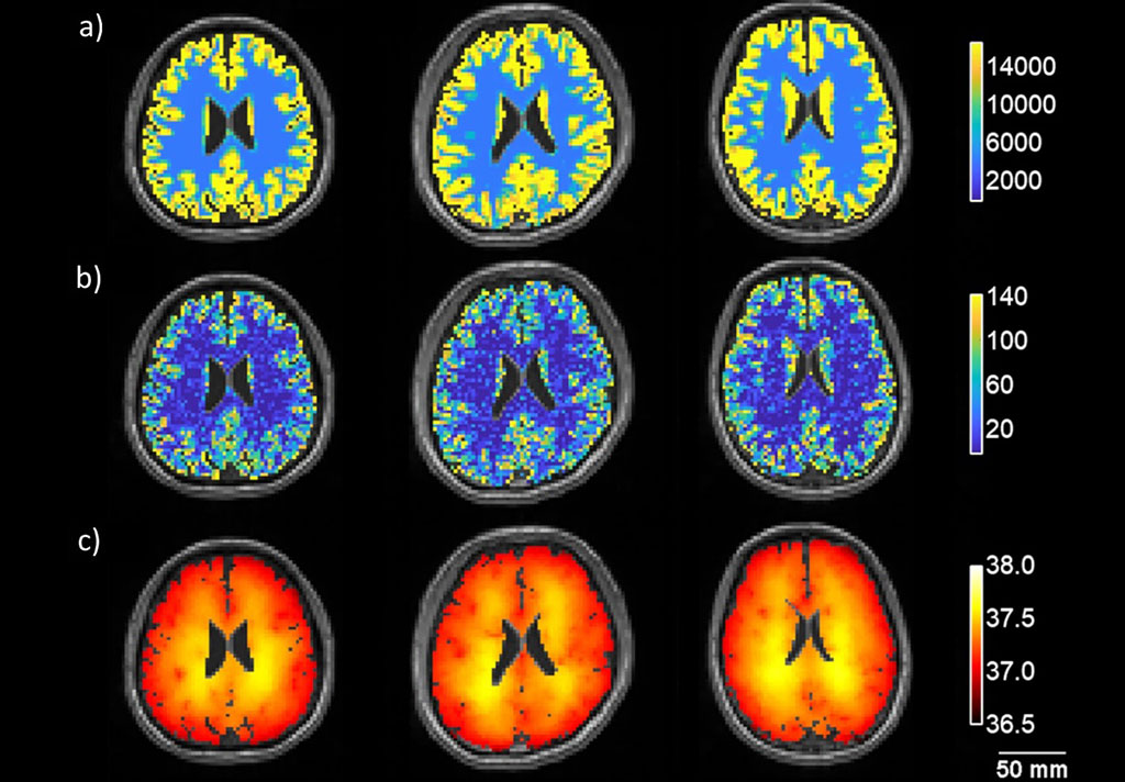
Developed by researchers at the Georgia Institute of Technology (Georgia Tech; Atlanta, GA, USA) and Emory University (Atlanta, GA, USA), the biophysical model can be used to generate 3D thermal distribution maps using whole brain MRI-based thermometry data. The model relies on two fundamental physical principles, conservation of energy and conservation of mass, to predict how heat is generated through metabolism and dissipated via blood flow throughout the brain, via conduction, convection, and advection.

Image: Metabolic heat (a), cerebral blood flow (b) and model-predicted brain temperature (c) in three volunteers (Photo courtesy of Emory University)
For the study, the researchers first collected structural MRI brain images from three healthy volunteers. After several hours of data analysis and simulations, the biophysical model computed the distribution of brain temperature in the brain, visualized as three-dimensional (3D) maps. To validate the model, the researchers then collected temperature measurements using whole-brain MRI thermometry, compared these measurements to the model’s predictions, and found that they agreed. The study was published on April 15, 2021, in Communications Physics.
“Brain temperature varies throughout the brain as blood flow and metabolic demands change. This means that you cannot assign a single temperature to a brain,” said co-senior author Andrei Fedorov, PhD, of Georgia Tech. “All of the complexity [in the brain’s functioning] results in a very unique personal temperature map that actually tells us a lot about what may be happening to us, what may have happened in the past, or what may happen if we somehow perturb us as a human.”
Despite being only a fraction of the human body’s mass, the brain accounts for 20% of total oxygen consumption in resting conditions, which is used for neuronal metabolism, neural processes, and the functioning of glial, endothelial, and epithelial cells. All this energy is transformed into intense heat production. The brain is also exceptionally well vascularized, receiving 15–20% of total cardiac output, which also serves as the means to remove the heat continuously generated through brain metabolism and neurological function.
Related Links:
Georgia Institute of Technology
Emory University


