A Minimal Footprint Magnetic Resonance Imaging (MRI) Scanner Helps Radiology Departments Address Overloads and Backlog
By MedImaging International staff writers
Posted on 23 Mar 2021
The Esaote (Genova, Italy) O-scan Elite MRI facilitates transversal, sagittal, coronal and oblique section imaging of the arm, including hand, wrist, forearm, and elbow, but excluding the upper part of the arm; and portions of the leg, including foot, ankle, calf, and knee, but excluding the thigh. As the O-scan MRI runs on a standard power outlet, and does not require helium cooling, it is a viable, cost effective solution that also impacts patient comfort. For pathologies in which it can be beneficial to image the joint in real-time movement, the Esaote True Motion can perform a regular MRI sequence. Posted on 23 Mar 2021
The Elite configuration comes with the Esaote AgilExam AI software, which recognizes the anatomy and automatically suggests the setup of the MRI scan according to the chosen protocol, improving image consistency and reducing exam times. Once the scout scan has been acquired, setup time for a complete exam is a matter of seconds, during which AgilExam adjusts the slice orientation and number of slices of all the scans, as per the chosen protocol. AgilExam is available for the knee, ankle, and wrist, but is not compatible with MRI conditional implants.
")
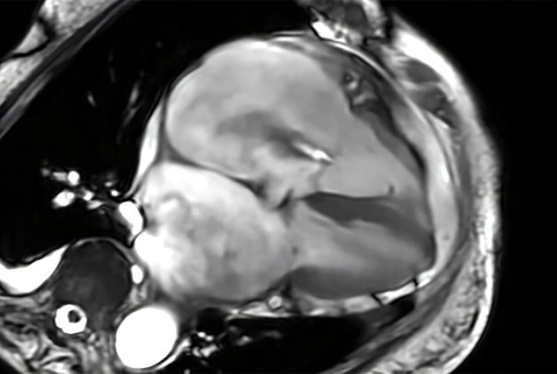
Image: The O-scan Elite dedicated MRI for extremities (Photo courtesy of Esaote)
“The Esaote Extremity MRI creates excellent diagnostic images of the elbow, wrist, hand, and fingers from the upper extremity and the knee, ankle, foot and toes from the lower extremity,” said Eric Lazar, MD, of University Radiology (East Brunswick, NJ, USA). “The patient reclines comfortably in an open-air environment while only the imaged body part lies within the machine. This is the first of its kind through our suite of 23 offices, and we are happy to welcome patients to our East Brunswick flagship office for this new service.”
MRI scanners can have ultraweak, weak, medium, strong, and superstrong magnetic fields, as measured in Tesla units. Highest-quality scans are usually taken with the aid of superconducting magnetic systems that generate very strong magnetic fields, providing the highest image resolution. But such high-field systems require liquid helium to keep the superconducting magnets cool, which demands high-power consumption, large separate facilities, and improved shielding.

Digital Intelligent Ferromagnetic Detector
Digital Ferromagnetic Detector

High-Precision QA Tool
DEXA Phantom

Diagnostic Ultrasound System
DC-80A

Ultrasound Needle Guidance System
SonoSite L25