Wearable MRI Detector Captures Anatomical Motion
By MedImaging International staff writers
Posted on 23 May 2018
A new study describes how a magnetic resonance imaging (MRI) element woven into a glove-like detector could aid the diagnosis of repetitive strain injuries such as carpal tunnel syndrome.Posted on 23 May 2018
Researchers at NYU Langone Medical Center (New York, NY, USA) and New York University (NYU, USA) have designed a wearable detector array for MRI of the hand that is based on high-impedance coils that can cloak themselves from electrodynamic interactions. The MRI signal is produced by hydrogen protons; since no electric current is created by the MRI signal, the new receiver coils no longer create magnetic fields that interfere with neighboring receivers, thus removing the need for rigid structures. The coils do not suffer from signal-to-noise (SNR) degradation mechanisms typically observed with the use of traditional low-impedance elements.

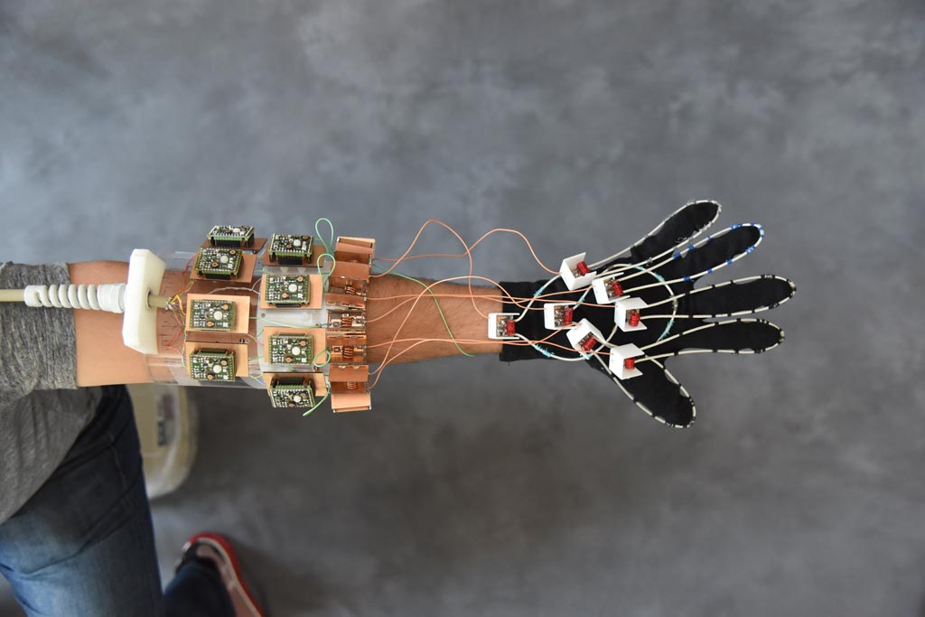
Image: The wearable MRI glove is designed to image moving joints and aid in diagnosing repetitive strain injuries (Photo courtesy of NYU Langone).
While MRI can efficiently image muscles, nerves, and even cartilage, which are difficult to study using other non-invasive methods, tendons and ligaments, which are made of dense proteins instead of fluid, remain difficult to see independently, because both appear as black bands running alongside bone. But with the new coils stitched into a cotton glove, they could generate images of freely moving muscles, tendons, and ligaments. The new coils revealed how the black bands moved in concert with the bones, which could help to catalogue differences that come with injury. The study was published on May 4, 2018, in Nature Biomedical Engineering.
“We wanted to try our new elements in an application that could never be done with traditional coils, and settled on an attempt to capture images with a glove,” said senior author Martijn Cloos, PhD, of the department of radiology at NYU Langone Health. “We hope that this result ushers in a new era of MRI design, perhaps including flexible sleeve arrays around injured knees, or comfy beanies to study the developing brains of newborns.”
The densely packed resonant structures used for MRI, such as nuclear magnetic resonance phased array detectors, suffer from resonant inductive coupling, which restricts the coil design to fixed geometries, in which receiver coils are painstakingly arranged to cancel out magnetic fields in neighboring coils. Once the best arrangement is set, the coils can no longer move relative to one another, constraining the ability of MRI to image complex, moving joints. But by using high-impedance detectors, the receiver coils no longer create magnetic fields that interfere with neighboring receivers, thus removing the need for rigid structures.
Related Links:
NYU Langone Medical Center
New York University


