MRI Multitasking Increases Diagnostic Accuracy and Reliability
By MedImaging International staff writers
Posted on 24 Apr 2018
A new cardiac magnetic resonance (CMR) imaging technique improves patient comfort and shortens testing time, according to a new study.Posted on 24 Apr 2018
Researchers at Cedars-Sinai Medical Center (Los Angeles, CA, USA), the University of California, Los Angeles (UCLA; USA), and Xuanwu Hospital (Beijing, China) conducted a study to examine how the need to reduce CMR imaging artefacts arising from body motion, the beating heart, and blood flow during quantitative imaging could be circumvented in order to make the procedure more reliable. The researchers decided therefore that rather than try to avoid the motion caused by breathing and heartbeats, they would embrace it.

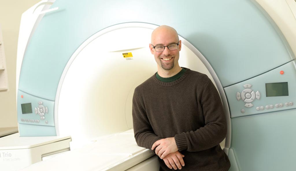
Image: Lead author Anthony Christodoulou, PhD (Photo courtesy of Anthony Christodoulou).
The new technique, which they dubbed CMR Multitasking, abstracts physiological motion and other dynamic processes as time extents, which can be resolved via low-rank tensor imaging, allowing for motion-resolved quantitative CMR in up to four time dimensions. The continuous-acquisition approach, captures--rather than avoids--motion, relaxation, and other dynamics, allowing for T1 mapping, T1/T2 mapping and time-resolved T1mapping of myocardial perfusion without electrocardiography (ECG) information and/or under free-breathing conditions. The study was published on April 9, 2018, in Nature Biomedical Engineering.
“MR Multitasking continuously acquires image data and then, when the test is completed, the program separates out the overlapping sources of motion and other changes into multiple time dimensions,” said lead author Anthony Christodoulou, PhD, of the Cedars-Sinai Biomedical Imaging Research Institute. “If a picture is 2D, then a video is 3D because it adds the passage of time. Our videos are 6D because we can play them back four different ways: We can playback cardiac motion, respiratory motion, and two different tissue processes that reveal cardiac health.”
“It is challenging to obtain good cardiac magnetic resonance images, because the heart is beating incessantly, and the patient is breathing, so the motion makes the test vulnerable to errors,” said Professor Shlomo Melmed, MB, ChB, dean of the Cedars-Sinai medical faculty. “By novel approaches to this longstanding problem, this research team has found a unique solution to improve cardiac care for patients around the world for years to come.”
CMR is a medical imaging technology for the non-invasive assessment of the function and structure of the cardiovascular system, based on the same basic principles as magnetic resonance imaging (MRI), with optimizations that use rapid imaging sequences. As a result, CMR images are currently acquired in steps. Patients breathe in and then hold their breath for each image, then recover before repeating the process for the next image.
Related Links:
Cedars-Sinai Medical Center
University of California, Los Angeles
Xuanwu Hospital


