Visual System Changes May Provide Biomarkers for PD
By MedImaging International staff writers
Posted on 10 Jul 2017
Researchers have found that visual system changes may provide an early indication of Parkinson’s disease and help clinicians monitor the disease.Posted on 10 Jul 2017
Such changes could become apparent ten years before the motor symptoms of the disease, and may include a change in visual acuity, an inability to perceive colors, and a decrease in the rate of blinking. Visual symptoms may appear more than 10 years before motor symptoms.

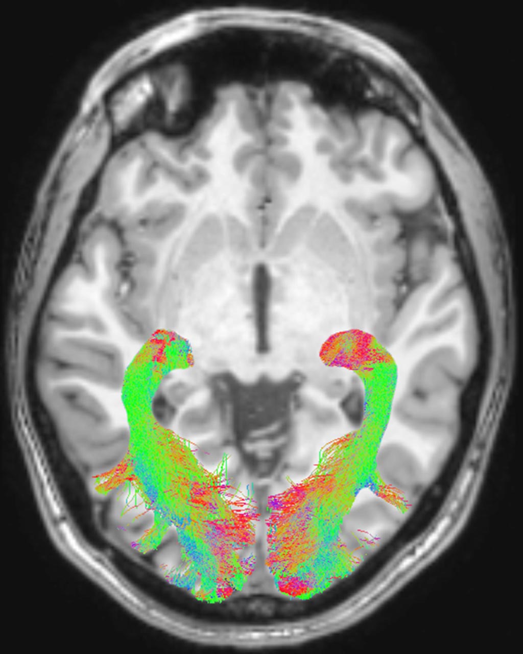
Image: The left and right Optic Radiation (OR) overlaid on a T1-weighted axial volume image of a subject (Photo courtesy of RSNA).
The researchers from the University Vita-Salute San Raffaele (Milan, Italy) published the results of their research online in the July 11, 2017, issue of the journal Radiology. The study included 11 men and 9 women, all newly diagnosed, but not yet treated for Parkinson’s disease (PD), and age- and gender-matched healthy control group of 20 people. The research team included ophthalmology, neurology and neuroradiology specialists.
The researchers used a Magnetic Resonance Imaging (MRI) Diffusion Weighted Imaging (DWI) to scan both the patients and the healthy controls. The patients were scanned within four weeks of their diagnosis to assess changes in the grey and white matter concentration of the brain, as well as Voxel-Based Morphometry (VBM). All participants of the study also underwent ophthalmologic examinations.
The results indicated significant abnormalities in the visual system brain structures of the Parkinson’s disease patients including a reduction of white matter concentration, alterations of optic radiations, and less optic chiasm volume.
Lead researcher, Dr. Alessandro Arrigo, MD, a resident in ophthalmology, said, "Although Parkinson’s disease is primarily considered a motor disorder, several studies have shown non-motor symptoms are common across all stages of the disease. These non-motor Parkinson’s symptoms may precede the appearance of motor signs by more than a decade. The study in depth of visual symptoms may provide sensitive markers of Parkinson’s disease. Visual processing metrics may prove helpful in differentiating Parkinsonism disorders, following disease progression, and monitoring patient response to drug treatment."
Related Links:
University Vita-Salute San Raffaele


