Brain Abnormalities Discovered in Bipolar Patients
By MedImaging International staff writers
Posted on 10 May 2017
The results of a new global MRI study show thinning of gray matter in patients with bipolar disorder.Posted on 10 May 2017
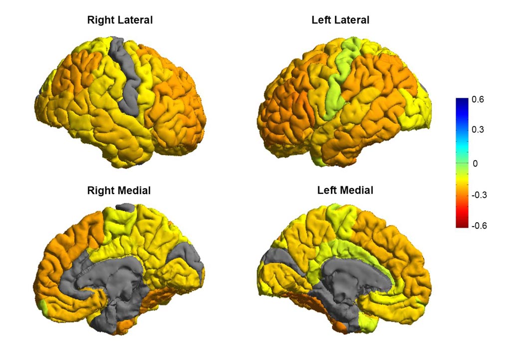
The researchers used the Magnetic Resonance Imaging (MRI) data from their study to build a roadmap of bipolar disorder, and the affects this condition has on the brain. The brain regions most affected by the disorder are used for inhibition, and emotions.

Image: Research shows bipolar patients tend to have gray matter reductions in frontal brain regions involved in self-control (orange colors), while sensory and visual regions are normal (gray colors) (Photo courtesy of the ENIGMA Bipolar Consortium/Derrek Hibar et al.).
The research was part of the international consortium ENIGMA (Enhancing Neuro Imaging Genetics Through Meta Analysis), and was led by the University of Southern California Keck School of Medicine Stevens Neuroimaging and Informatics Institute. The research findings were published in the May 2, 2017, issue of the journal Molecular Psychiatry. For their study, the researchers used MRI scans of 2,447 adults suffering from bipolar disorder, as well as scans from 4,056 healthy control subjects.
The results of the study showed thinning of brain gray matter in patients with the disorder, compared to the healthy control subjects. The researchers found that the greatest deficits were in the frontal and temporal regions of the brain controlling inhibition and motivation.
According to the researchers, the clear and consistent changes in key brain regions provide insight into the underlying mechanisms of the disorder, and could enable future research into new medications and treatments.
Senior author of the study, Ole A. Andreassen, professor, Norwegian Centre for Mental Disorders Research, University of Oslo, said, "We created the first global map of bipolar disorder and how it affects the brain, resolving years of uncertainty on how people's brains differ when they have this severe illness."


