MRI Reveals Changes in Brains of Pregnant Women
By MedImaging International staff writers
Posted on 12 Jan 2017
Magnetic resonance imaging (MRI) scans reveals that the brains of pregnant women show significant reductions in grey matter in regions associated with social cognition.Posted on 12 Jan 2017
Researchers at Universitat Autònoma de Barcelona, Fundació IMIM, Leiden University, and other institutions conducted a study that compared MRI scans of 25 first-time mothers before and after their pregnancy, of 19 of their male partners, and of a control group formed by 20 women who were not and had never been pregnant and 17 of their male partners. The data was gathered during a period of five years and four months.

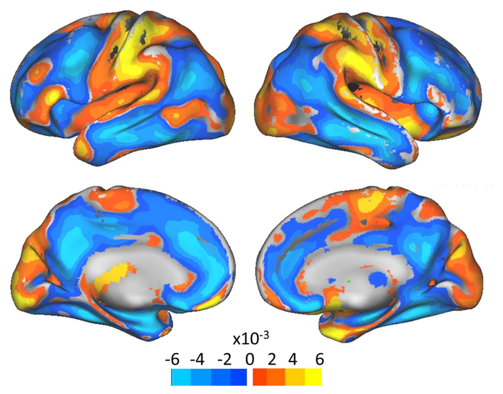
Image: Grey matter reduction in pregnant women (orange), compared to controls (Photo courtesy of UAB).
The results of the study showed a symmetrical reduction in the volume of grey matter in the medial frontal and posterior cortex line, as well as in specific sections of, mainly, prefrontal and temporal cortex in pregnant women. The reductions in grey matter were practically identical variations in both women who underwent fertility treatments and women who became pregnant naturally. In MRI scans taken two years later, the gray matter loss remained, except in the hippocampus, where most volume had been restored.
The changes were so consistent that a computer algorithm could predict with 100% accuracy whether a woman had been pregnant from her MRI scan. According to the researchers, the grey matter areas corresponded with a neural network that is associated with processes involved in social cognition and self-focused processing, with a similar decline in gray matter volume occurring during adolescence. The study was published on December 19, 2016, in Nature Neuroscience.
“We certainly don’t want to put a message out there along the lines of ‘pregnancy makes you lose your brain’. Gray matter volume loss can also represent a beneficial process of maturation or specialization, an adaptive process of functional specialization towards motherhood,” said lead author neuroscientist Elseline Hoekzema, PhD, of UAB and Leiden University. “These changes may reflect, at least in part, a mechanism of synaptic pruning, where weak synapses are eliminated giving way to more efficient and specialized neural networks.”
“The loss of grey matter does not imply any cognitive deficits, but rather points to an adaptive process related to the benefits of better detecting the needs of the child, such as identifying the newborn's emotional state,” added senior author Oscar Vilarroya, PhD, of the UAB cognitive neuroscience research unit. “Moreover, they provide primary clues regarding the neural basis of motherhood, perinatal mental health, and brain plasticity in general.”


